. 1999 Aug 3;96(16):9021-6.
doi: 10.1073/pnas.96.16.9021.

Focal adhesion kinase promotes phospholipase C-gamma1 activity

Affiliations

Focal adhesion kinase promotes phospholipase C-gamma1 activity

X Zhang et al. Proc Natl Acad Sci U S A. .

Abstract

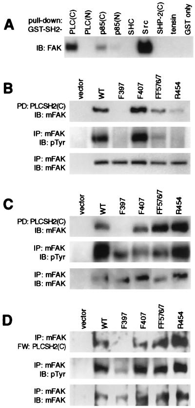
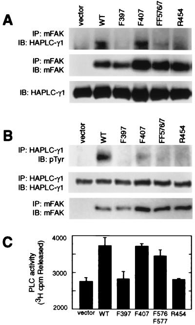
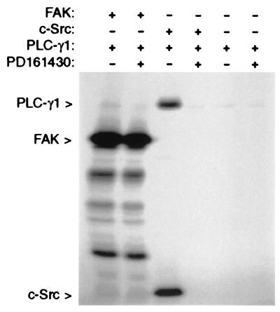
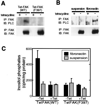
The nonreceptor tyrosine kinase FAK ("focal adhesion kinase") is a key mediator of integrin signaling events controlling cellular responses to the extracellular matrix, including spreading, migration, proliferation, and survival. Integrin-ligand interactions stimulate FAK tyrosine phosphorylation and activation of FAK signaling functions. Here evidence is presented that the FAK autophosphorylation site Tyr-397 mediates a direct interaction with the C-terminal Src homology 2 domain of phospholipase C (PLC)-gamma1 and that this is required for both adhesion-dependent association of the two molecules and increased inositol phosphate production in mouse embryo fibroblasts. Overexpression of FAK and PLC-gamma1 in COS-7 cells increases PLC-gamma1 enzymatic activity and tyrosine phosphorylation, also dependent on FAK Tyr-397. However, FAK appears incapable of directly phosphorylating PLC-gamma1. These observations suggest a role for FAK in recruiting PLC-gamma1 to the plasma membrane at sites of cell-matrix adhesion and there promoting its enzymatic activity, possibly by releasing the repression caused by intramolecular interactions of the PLC-gamma1 Src homology domains and/or by positioning it for phosphorylation by associated Src-family kinases. These findings expand the known signaling functions of FAK and provide mechanistic insight into integrin-stimulation of PLC-gamma1.


Figures

Figure 1
Figure 1
The C-terminal SH2 domain of PLC-γ1 interacts with FAK pTyr397. (A) Lysates of adherent BALB/c-3T3 cells were incubated with various GST-SH2 proteins (or GST only) immobilized on glutathione agarose beads. Proteins “pulled down” by the beads were separated by SDS/PAGE and were immunoblotted (IB) with anti-FAK polyclonal antibody 331. (B) Lysates of COS-7 cells expressing Myc-tagged FAK (mFAK) variants (WT, F397, F407, FF576/7, or R454) were subjected to GST-PLC-γ1-SH2C pull-down [PD: PLCSH2(C)], and the presence of mFAK variants was detected by immunoblotting with anti-Myc antibody 9E10 (Top). Relative mFAK protein and pTyr levels in the lysates were determined by immunoprecipitation with 9E10 followed by immunoblotting with either 9E10 (Bottom) or anti-pTyr antibody 4G10 (Middle), respectively. (C) mFAK variants were coexpressed with activated Src in COS-7 cells, and lysates were examined for their association with GST-PLC-γ1-SH2C (Top), tyrosine phosphorylation (Middle), and protein levels (Bottom) as above. (D) mFAK variants coexpressed with active Src in COS-7 cells were isolated by immunoprecipitation using 9E10 antibody and were assessed for direct binding of GST-PLC-γ1-SH2C by Far-Western (FW) analysis (Top). The membrane was sequentially stripped and reprobed with 4G10 (Middle) and 9E10 (Bottom) antibodies to determine relative mFAK pTyr and protein levels, respectively. The FAK expression vector alone was used as control for assays in B, C, and D.
Figure 2
Figure 2
FAK associates with PLC-γ1 and stimulates PLC-γ1 tyrosine phosphorylation and enzymatic activity in COS-7 cells. (A) Myc-tagged FAK (mFAK) variants (or FAK vector only control) were transiently coexpressed in COS-7 cells together with HA-tagged PLC-γ1, and mFAK variants were immunoprecipitated (IP) with antibody 9E10. Coprecipitation of HA-PLC-γ1 was assessed by probing immunoblots (IB) with anti-HA antibody 12CA5 (Top), and relative amounts of immunoprecipitating mFAK were visualized by immunoblotting with 9E10 (Middle). Expression of HA-PLC-γ1 was assessed by immunoblot analysis of whole cell lysates using 12CA5 (Bottom). (B) HA-PLC-γ1 coexpressed in COS-7 cells with mFAK variants (or vector only) was immunoprecipitated with 12CA5 antibody and was analyzed for pTyr content by immunoblot analysis using anti-pTyr antibody 4G10 (Top). Recovery of HA-PLC-γ1 was determined by probing immunoblots with 12CA5 antibody (Middle), and expression of mFAK was determined by immunoprecipitation/immunoblot analysis using 9E10 antibody (Bottom). (C) HA-PLC-γ1 coexpressed in COS-7 cells with mFAK variants (or vector only) was immunoprecipitated with 12CA5 antibody and was analyzed for hydrolytic activity toward [3H]phosphatidylinositol 4,5-bisphosphate. PLC-γ1 activity is presented as cpm [3H]inositol 1,4,5-trisphosphate released (mean values + SEM from four independent experiments). Separate immunoblots (not shown) confirmed that near-equal amounts of HA-PLC-γ1 were assayed under each condition and that near-equal amounts of mFAK variants were coexpressed.
Figure 3
Figure 3
FAK does not phosphorylate PLC-γ1 in vitro. Recombinant PLC-γ1 was incubated either alone or in the presence of recombinant FAK or recombinant c-Src in a kinase assay reaction mixture containing [γ-32P]ATP. The Src-selective inhibitor PD16430 was included in some reactions to guard against possible contamination of the FAK or PLC-γ1 preparations with Src-family kinases. After the reactions, proteins were separated by SDS/PAGE, and [32P]-labeled proteins were detected by autoradiography.
Figure 4
Figure 4
FAK Tyr397 is required for cell adhesion-promoted FAK:PLC-γ1 association and inositol phosphate production. (A) TetFAK(WT) and TetFAK(F397) cells maintained in the presence or absence of tetracycline were lysed at subconfluent density, and FAK immunoprecipitates (IP) were examined for the presence of PLC-γ1 (Upper) or FAK (Lower) by immunoblot (IB) analysis. (B) The same cells, maintained in the presence or absence of tetracycline, were serum-starved (20 hr in DMEM containing 0.5% FBS) and then were detached by trypsin and either were maintained in suspension or were replated onto fibronectin-coated dishes and allowed to spread for 30 min. Both suspended and fibronectin-replated cells then were lysed, and FAK immunoprecipitates were subjected to immunoblot analysis using antibodies against either PLC-γ1 (Upper) or FAK (Lower). (C) Serum- and inositol-starved adherent TetFAK(WT) and TetFAK(F397) cells were labeled with myo-(2-3H) inositol, were harvested by trypsinization, and were resuspended in serum- and inositol-free DMEM. One half of the cell suspension was replated onto a fibronectin-coated cell culture dish and was allowed to attach and spread whereas the other half was held in suspension. After 30 min, free inositol phosphates of both suspended and fibronectin-adherent cells were extracted, and [3H]inositol phosphates were quantitated by scintillation counting. The relative production of total inositol phosphates is shown as cpm per mg of total residual cellular protein. Data presented are means (+SEM) from four independent measurements.

References

    1. Rosales C, O’Brien V, Kornberg L, Juliano R. Biochim Biophys Acta. 1995;1242:77–98. - PubMed
    1. Schwartz M A, Schaller M D, Ginsberg M H. Annu Rev Cell Dev Biol. 1995;11:549–599. - PubMed
    1. Hanks S K, Polte T R. BioEssays. 1997;19:137–145. - PubMed
    1. Schaller M D, Borgman C A, Cobb B S, Vines R R, Reynolds A B, Parsons J T. Proc Natl Acad Sci USA. 1992;89:5192–5196. - PMC - PubMed
    1. Hanks S K, Calalb M B, Harper M C, Patel S K. Proc Natl Acad Sci USA. 1992;89:8487–8491. - PMC - PubMed

Publication types

MeSH terms

LinkOut - more resources