. 1999 Aug 3;96(16):9083-8.
doi: 10.1073/pnas.96.16.9083.

MSE55, a Cdc42 effector protein, induces long cellular extensions in fibroblasts

Affiliations

MSE55, a Cdc42 effector protein, induces long cellular extensions in fibroblasts

P D Burbelo et al. Proc Natl Acad Sci U S A. .

Abstract

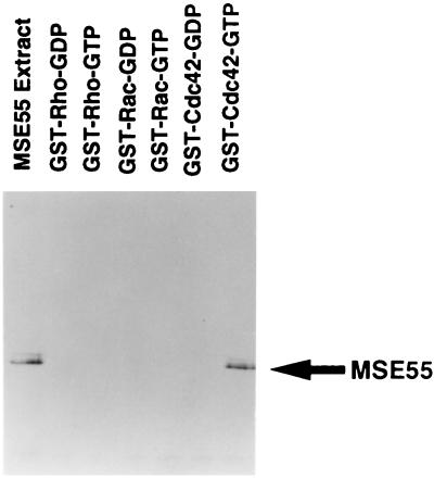
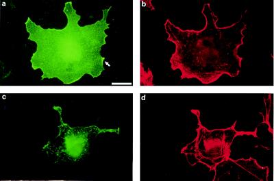
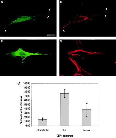
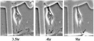
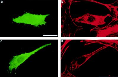
Cdc42 is a member of the Rho GTPase family that regulates multiple cellular activities, including actin polymerization, kinase-signaling activation, and cell polarization. MSE55 is a nonkinase CRIB (Cdc42/Rac interactive-binding) domain-containing molecule of unknown function. Using glutathione S-transferase-capture experiments, we show that MSE55 binds to Cdc42 in a GTP-dependent manner. MSE55 binding to Cdc42 required an intact CRIB domain, because a MSE55 CRIB domain mutant no longer interacted with Cdc42. To study the function of MSE55 we transfected either wild-type MSE55 or a MSE55 CRIB mutant into mammalian cells. In Cos-7 cells, wild-type MSE55 localized at membrane ruffles and increased membrane actin polymerization, whereas expression of the MSE55 CRIB mutant showed fewer membrane ruffles. In contrast to these results, MSE55 induced the formation of long, actin-based protrusions in NIH 3T3 cells as detected by immunofluorescence and live-cell video microscopy. MSE55-induced protrusion formation was blocked by expression of dominant-negative N17Cdc42, but not by expression of dominant-negative N17Rac. These findings indicate that MSE55 is a Cdc42 effector protein that mediates actin cytoskeleton reorganization at the plasma membrane.


Figures

Figure 1
Figure 1
MSE55 interacts with Cdc42 in a GTP-dependent fashion. Cos-1 cells were transfected with FLAG epitope-tagged MSE55 eukaryotic expression vector. Cell lysates obtained from these cells were incubated with glutathione S-transferase-bound Rho, Rac, and Cdc42 that had been preloaded with either GDP or guanosine 5′-[γ-thio]triphosphate. After four washes, bound proteins were eluted and analyzed by Western blotting with M2 anti-FLAG mAb and goat anti-mouse-horseradish peroxidase and detected by enhanced chemiluminescence. An aliquot of cell lysate from FLAG-MSE55-transfected cells also is shown.
Figure 2
Figure 2
MSE55 localizes to lamellipodia in Cos-7 cells. FLAG-tagged MSE55 was transfected into Cos-7 cells. Expression of wild-type MSE55 (a) or a CRIB mutant, MSE55-D36A, P41A, H47A (c), was detected with the M2 anti-FLAG mouse mAb and goat anti-mouse IgG-FITC. Staining of F-actin filaments was visualized with Texas Red-phalloidin (b and d). Fluorescent micrographs show MSE55 staining in membrane ruffles 18 hr after transfection (a and b). MSE55-D36A, P41A, H47A-expressing cells (c) showed less staining in the ruffles and more stress fibers (d). (Bar = 20 μm.)
Figure 3
Figure 3
MSE55 induces long cellular extensions in NIH 3T3 cells. FLAG-tagged MSE55 was transfected into NIH 3T3 cells. Expression of wild-type MSE55 (a) or the MSE55-D36A, P41A, H47A CRIB mutant (c) was detected with M2 anti-FLAG mouse mAb and goat anti-mouse IgG-FITC. Staining of F-actin filaments also was visualized with Texas Red-phalloidin (b and d). Fluorescent micrographs show MSE55-expressing cells showing long cellular extensions (a) that colocalize with F-actin staining (b). Expression of MSE55-D36A, P41A, H47A did not show long cellular extensions (c and d). (Bar = 20 μm.) NIH 3T3 cells with extensions greater than 10 μm in length were counted in untransfected and both wild-type and mutant MSE55-transfected cells (e). Results are expressed as the mean percentage of cells (±SD) from three separate experiments.
Figure 4
Figure 4
MSE55 expression induces membrane protrusions. NIH 3T3 cells were seeded on gridded coverslips 24 hr before microinjection. Cells were comicroinjected with mammalian expression vectors for MSE55 and EGFP (used as a tracking agent). Upon detection of EGFP expression, time-lapse video microscopy was begun and digitized images were captured at 2-min intervals. Cell is shown at 3.5, 4, and 9 hr after microinjection.
Figure 5
Figure 5
Extension formation does not require Rac Activity. NIH 3T3 cells were cotransfected with MSE55 and dominant-negative Rac. N17Rac transfected cells were detected with rabbit polyclonal anti-Myc antibody and goat anti-rabbit IgG-FITC (green). N17Rac-expressing cells produce multiple thin filopodia (a and b). N17Rac, MSE55, and F-actin were detected by using triple immunofluorescence. Cells coexpressing N17Rac and MSE55 form multiple thin filopodia and additionally form a single, long cellular extension (c and d). MSE55 was detected with M2 anti-FLAG mouse mAb and goat anti-mouse IgG-Alexa 350 (data not shown). Expression of dominant-negative N17Rac (green) was detected with a rabbit polyclonal antibody against c-Myc and with anti-rabbit IgG-FITC. F-actin was detected with Texas Red-phalloidin (red). (Bar = 20 μm.)

References

    1. Stossel T P. Science. 1993;260:1086–1094. - PubMed
    1. Kozma R, Ahmed S, Best A, Lim L. Mol Cell Biol. 1995;15:1942–1952. - PMC - PubMed
    1. Nobes K, Hall A. Cell. 1995;81:53–62. - PubMed
    1. Ridley A J, Paterson H F, Johnston C L, Diekmann D, Hall A. Cell. 1992;70:401–410. - PubMed
    1. Ridley A J, Hall A. Cell. 1992;70:389–399. - PubMed

Publication types

MeSH terms

Associated data