

Isolation of primitive human hematopoietic progenitors on the basis of aldehyde dehydrogenase activity
- PMID: 10430905
- PMCID: PMC17742
- DOI: 10.1073/pnas.96.16.9118
Isolation of primitive human hematopoietic progenitors on the basis of aldehyde dehydrogenase activity
Abstract
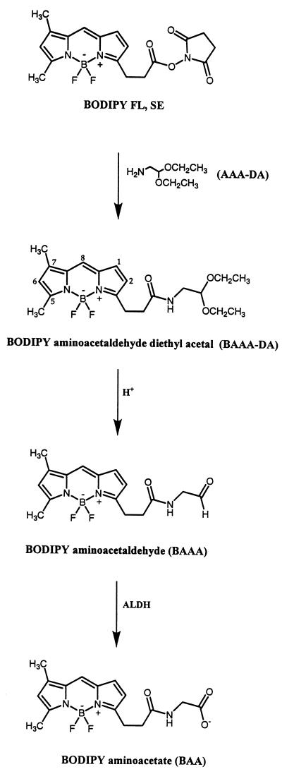
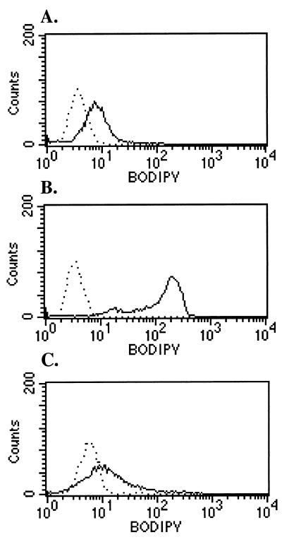
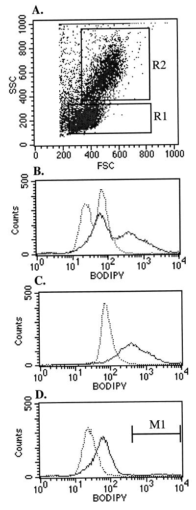
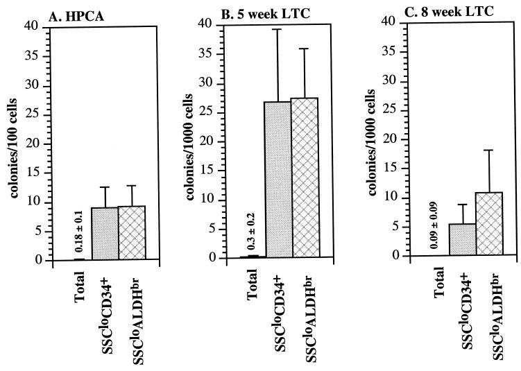
Because hematopoietic stem cells are rich in aldehyde dehydrogenase (ALDH) activity, we developed a fluorescent substrate for ALDH, termed BODIPY aminoacetaldehyde (BAAA), and tested its potential for isolating primitive human hematopoietic cells. A population of cells with low orthogonal light scattering and bright fluorescence intensity (SSC(lo)ALDH(br) cells) could be readily fractionated from human umbilical cord blood cells costained with BAAA and the multidrug-resistance inhibitor verapamil. The SSC(lo)ALDH(br) population was depleted of lineage-committed cells, 40-90% pure for CD34(+)CD38(lo/-) cells, and enriched 50- to 100-fold for primitive hematopoietic progenitors detected in short- and long-term culture analyses. Together, these observations indicate that fractionating human hematopoietic stem cells on the basis of ALDH activity using BAAA is an effective method for isolating primitive human hematopoietic progenitors. This technique may be useful for isolating stem cells from other tissues as well.
Figures

Similar articles
-
Isolation of early hematopoietic cells, including megakaryocyte progenitors, in the ALDH-bright cell population of cryopreserved, banked UC blood.Cytotherapy. 2007;9(6):569-76. doi: 10.1080/14653240701466347. Cytotherapy. 2007. PMID: 17882722
-
Distinct hematopoietic progenitor compartments are delineated by the expression of aldehyde dehydrogenase and CD34.Blood. 2005 Jul 1;106(1):95-102. doi: 10.1182/blood-2004-09-3652. Epub 2005 Mar 24. Blood. 2005. PMID: 15790790 Free PMC article.
-
Dual SP/ALDH functionalities refine the human hematopoietic Lin-CD34+CD38- stem/progenitor cell compartment.Stem Cells. 2009 Oct;27(10):2552-62. doi: 10.1002/stem.186. Stem Cells. 2009. PMID: 19650038
-
Next generation of ALDH substrates and their potential to study maturational lineage biology in stem and progenitor cells.Am J Physiol Gastrointest Liver Physiol. 2015 Apr 1;308(7):G573-8. doi: 10.1152/ajpgi.00420.2014. Epub 2015 Feb 5. Am J Physiol Gastrointest Liver Physiol. 2015. PMID: 25656041 Free PMC article. Review.
-
The Multifaceted Role of Aldehyde Dehydrogenases in Prostate Cancer Stem Cells.Cancers (Basel). 2021 Sep 20;13(18):4703. doi: 10.3390/cancers13184703. Cancers (Basel). 2021. PMID: 34572930 Free PMC article. Review.
Cited by
-
Dietary Glucose Consumption Promotes RALDH Activity in Small Intestinal CD103+CD11b+ Dendritic Cells.Front Immunol. 2020 Aug 11;11:1897. doi: 10.3389/fimmu.2020.01897. eCollection 2020. Front Immunol. 2020. PMID: 32849649 Free PMC article.
-
Sensitizing mucoepidermoid carcinomas to chemotherapy by targeted disruption of cancer stem cells.Oncotarget. 2016 Jul 5;7(27):42447-42460. doi: 10.18632/oncotarget.9884. Oncotarget. 2016. PMID: 27285758 Free PMC article.
-
Biological and clinical relevance of stem cells in pancreatic adenocarcinoma.J Gastroenterol Hepatol. 2012 Mar;27 Suppl 2(Suppl 2):15-8. doi: 10.1111/j.1440-1746.2011.07015.x. J Gastroenterol Hepatol. 2012. PMID: 22320910 Free PMC article. Review.
-
Targeting cancer stem cells in squamous cell carcinoma.Precis Clin Med. 2019 Sep;2(3):152-165. doi: 10.1093/pcmedi/pbz016. Epub 2019 Oct 1. Precis Clin Med. 2019. PMID: 31598386 Free PMC article. Review.
-
Alcohol abuse and disorder of granulopoiesis.Pharmacol Ther. 2019 Jun;198:206-219. doi: 10.1016/j.pharmthera.2019.03.001. Epub 2019 Mar 1. Pharmacol Ther. 2019. PMID: 30831129 Free PMC article. Review.
References
-
- Morrison S J, Wright D E, Cheshier S H, Weissman I L. Curr Opin Immunol. 1997;9:216–221. - PubMed
-
- Spangrude G, Weissman I. Science. 1988;241:58–62. - PubMed
-
- Civin C I, Strauss L, Brovall C, Fackler M, Schwartz J. J Immunol. 1984;133:157–165. - PubMed
-
- Krause D, Ito T, Fackler M, Smith O, Collector M, Sharkis S, Mary S. Blood. 1994;84:691–701. - PubMed
-
- Osawa M, Hanada K, Hamada H, Nakauchi H. Science. 1996;273:242–245. - PubMed
Publication types
MeSH terms
Substances
Grants and funding
LinkOut - more resources
Full Text Sources
Other Literature Sources
Medical
Research Materials
