. 1999 Aug 3;96(16):9142-7.
doi: 10.1073/pnas.96.16.9142.

A vital role for glycosphingolipid synthesis during development and differentiation

Affiliations

A vital role for glycosphingolipid synthesis during development and differentiation

T Yamashita et al. Proc Natl Acad Sci U S A. .

Abstract

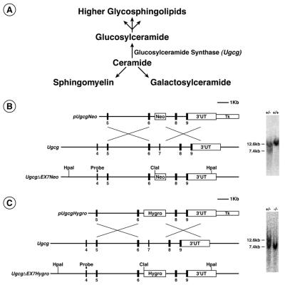
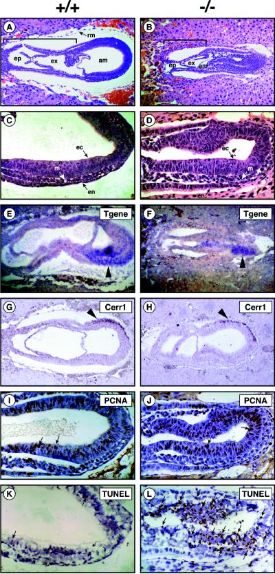
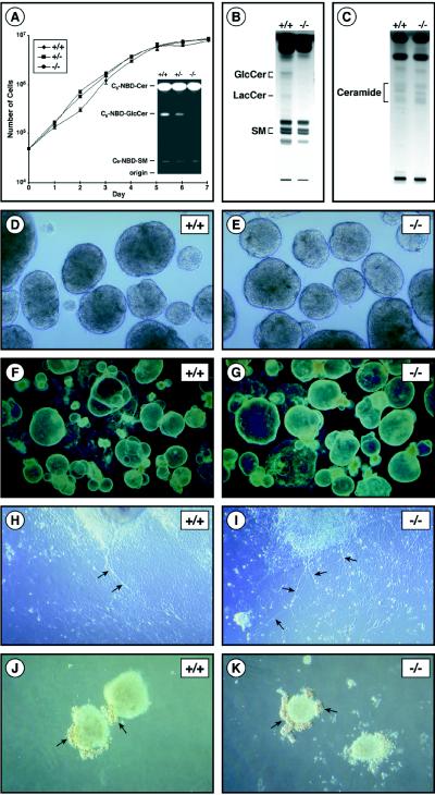
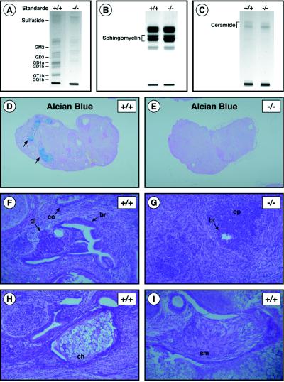
Glycosphingolipids (GSLs) are believed to be integral for the dynamics of many cell membrane events, including cellular interactions, signaling, and trafficking. We have investigated their roles in development and differentiation by eliminating the major synthesis pathway of GSLs through targeted disruption of the Ugcg gene encoding glucosylceramide synthase. In the absence of GSL synthesis, embryogenesis proceeded well into gastrulation with differentiation into primitive germ layers and patterning of the embryo but was abruptly halted by a major apoptotic process. In vivo, embryonic stem cells deficient in GSL synthesis were again able to differentiate into endodermal, mesodermal, and ectodermal derivatives but were strikingly deficient in their ability to form well differentiated tissues. In vitro, however, hematopoietic and neuronal differentiation could be induced. The results demonstrate that the synthesis of GSL structures is essential for embryonic development and for the differentiation of some tissues and support the concept that GSLs are involved in crucial cell interactions mediating these processes.


Figures

Figure 1
Figure 1
Ugcg gene targeting. (A) The biosynthetic pathways for sphingolipids. Glucosylceramide is produced by the modification of ceramide by glucosylceramide synthase. Glucosylceramide is the core structure for the synthesis of higher-order GSLs. Ceramide also is used for synthesis of sphingomyelin and galactosylceramide. (B) The structure of the pUgcgNeo targeting vector is shown at the top. The wild-type Ugcg locus is in the middle. The structure of the targeted locus (UgcgΔEX7Neo) is on the bottom. The UgcgΔEX7Neo locus was detected by Southern blot analysis of genomic DNA after digestion with HpaI and ClaI and hybridization with the probe indicated. The wild-type allele yielded a 12.6-kb fragment, and the UgcgΔEX7Neo allele yielded a 7.4-kb fragment. The Southern blots were from tail DNA of wild-type (+/+), and UgcgΔEX7Neo/+ (+/−) mice derived from targeted ES cells. (C) The structure of the pUgcgHygro targeting vector is shown at the top. The wild-type Ugcg locus is in the middle. The structure of the targeted allele, UgcgEX7Hygro, is on the bottom. The UgcgEX7Hygro locus was detected by Southern blot analysis of genomic DNA after digestion with HpaI and ClaI and hybridization with the probe indicated. The wild-type allele yielded a 12.6-kb band, and the UgcgΔEX7Neo and UgcgΔEX7Hygro alleles each yielded a 7.4-kb fragment. The Southern blots were of DNA of UgcgΔEX7Neo (+/−) ES cells and doubly targeted ES cells UgcgΔEX7Neo/ΔEX7Hygro (−/−).
Figure 2
Figure 2
Histology of mutant Ugcg embryos. E7.5 embryos were obtained from timed matings between UgcgΔEX7Neo/+ males and UgcgΔEX7Neo/+ females. A, C, E, G, I, and K show wild-type embryos (+/+). B, D, F, H, J, and L show UgcgΔEX7Neo/ΔEX7Neo embryos (−/−). (A and B) Comparison of the organization of embryos by H & E staining of paraffin sections. The cavities are indicated. Note the smaller size and disorganization of the mutant embryo (×100). The bracket delineates the extraembryonic region. ep, ectoplacental cavity; ex, exoceolom; am, amniotic cavity; rm, Reichert’s membrane. (C and D) Morphologic demonstration of embryonic ectoderm (ec), mesoderm (m), and endoderm (en) in H & E stained paraffin sections of wild-type and mutant embryos (×250). (E and F) In situ hybridization using a Brachyury (T gene) probe. T gene hybridization signal (arrow head) was detected in both wild-type and mutant embryos (×100). (G and H) In situ hybridization using Cerr1 probe. Cerr1 hybridization signal (arrow head) was detected in anterior endoderm of both wild-type and mutant embryos (×100). (I and J) Immunodectection of PCNA. A similar level of expression (dark brown stain) is seen in the wild-type and mutant embryo. Arrows point out some examples of positive cells (×250). (K and L) TUNEL detection of apoptotic cells. Greatly enhanced incidence of TUNEL-positive nuclei (small, dark brown spots) was seen in the mutant embryo, especially in the ectodermal layer. Arrows point to some of the many positive reactions in the mutant embryo. Only a few positive reactions were found in the wild-type embryo (×250).
Figure 3
Figure 3
In vitro growth and differentiation of ES cells in the absence of GSL synthesis. (A Left) Growth of wild-type (+/+), UgcgΔEX7Neo/+ (+/−), and UgcgΔEX7Neo/ΔEX7Hygro (−/−) ES cells. ES cells (5 × 10⁴) were seeded into 60-mm plates containing mitomycin C-treated embryonic fibroblasts to initiate the experiment. Each day the medium was changed and the cells from one plate were harvested and were counted. (Inset) Glucosylceramide synthase activity in wild-type (+/+), UgcgΔEX7Neo/+ (+/−), and UgcgΔEX7Neo/ΔEX7Hygro (−/−) ES cells. Each cell extract, corresponding to 50 μg of protein, was incubated with C6-NBD-ceramide and UDP-glucose. After incubation, lipids were separated by high-performance thin-layer chromatography (HPTLC) and were visualized by UV illumination. The positions of the C6-NBD derivatives of glucosylceramide (C6-NBD-GlcCer) lactosylceramide (C6-NBD-LacCer) and sphingomyelin (C6-NBD-SM) are indicated (B) HPTLC analysis of neutral glycolipids isolated from wild-type (+/+) and UgcgΔEX7Neo/ΔEX7Hygro (−/−) ES cells. The positions of glucosylceramide (GlcCer), lactosylceramide (LacCer), and sphingomyelin (SM) are indicated. (C) HPTLC analysis of ceramide levels in wild-type (+/+) and UgcgΔEX7Neo/ΔEX7Hygro (−/−) ES cells. The position of ceramide is indicated. (D and E) Wild-type (+/+) and UgcgΔEX7Neo/ΔEX7Hygro (−/−) embryoid bodies after 3 days of culture (×60). (F and G) Wild-type (+/+) and UgcgΔEX7Neo/ΔEX7Hygro (−/−) cystic embryoid bodies after 22 days of culture (×20). (H and I) Retinoic acid-induced neuronal differentiation of wild-type (+/+) and UgcgΔEX7Neo/ΔEX7Hygro (−/−) ES cells (×100). Arrows indicate neurite formation. (J and K) Erythropoietin-induced hematopoietic differentiation of wild-type (+/+) and UgcgΔEX7Neo/ΔEX7Hygro (−/−) ES cells (×100). Arrows indicate clusters of red erythroid cells surrounding embryoid bodies.
Figure 4
Figure 4
In vivo differentiation of teratomas in the absence of GSL synthesis. (A) Acidic lipid fractions from of wild-type (+/+) and UgcgΔEX7Neo/ΔEX7Hygro (−/−) teratomas analyzed by HPTLC. The position of GSL standards are indicated on the left. (B) HPTLC analysis of sphingomyelin levels from of wild-type (+/+) and UgcgΔEX7Neo/ΔEX7Hygro (−/−) teratomas. The position of sphingomyelin is indicated. (C) HPTLC analysis of ceramide levels in wild-type (+/+) and UgcgΔEX7Neo/ΔEX7Hygro (−/−) teratomas. The position of ceramide is indicated. (D and E) Sections of wild-type (+/+) and UgcgΔEX7Neo/ΔEX7Hygro (−/−) teratomas stained with Alcian blue (×5). Arrows indicate Alcian blue-positive cartilage in the wild-type tumor but not found in the mutant tumor. (F) H & E-stained section of the wild-type teratoma showing well differentiated epithelial tissues (×100). gl, glands; br, bronchial epithelium; co, colonic epithelium. (G) H & E-stained section of UgcgΔEX7Neo/ΔEX7Hydro (−/−) teratoma showing poorly differentiated epithelial tissue (ep) and small focus of bronchial epithelium (br) (×100). (H) H & E-stained section of a wild-type teratoma showing chondrocytes (ch) (×100). (I) H & E-stained section of the wild-type teratoma showing smooth muscle (sm) (×100).

References

    1. Svennerholm L. Prog Brain Res. 1994;101:x–xiv. - PubMed
    1. Kolter T, Sandhoff K. Brain Pathol. 1998;8:79–100. - PMC - PubMed
    1. Hakomori S. J Biol Chem. 1990;265:18713–18716. - PubMed
    1. Hakomori S, Igarashi Y. J Biochem (Tokyo) 1995;118:1091–1103. - PubMed
    1. Nagai Y, Tsuji S. Prog Brain Res. 1994;101:119–126. - PubMed

Publication types

MeSH terms