. 1999 Aug 3;96(16):9236-41.
doi: 10.1073/pnas.96.16.9236.

Digital PCR

Affiliations

Digital PCR

B Vogelstein et al. Proc Natl Acad Sci U S A. .

Abstract

The identification of predefined mutations expected to be present in a minor fraction of a cell population is important for a variety of basic research and clinical applications. Here, we describe an approach for transforming the exponential, analog nature of the PCR into a linear, digital signal suitable for this purpose. Single molecules are isolated by dilution and individually amplified by PCR; each product is then analyzed separately for the presence of mutations by using fluorescent probes. The feasibility of the approach is demonstrated through the detection of a mutant ras oncogene in the stool of patients with colorectal cancer. The process provides a reliable and quantitative measure of the proportion of variant sequences within a DNA sample.


Figures
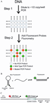
Figure 1
Figure 1
Schematic of Dig-PCR. (A) The basic two steps involved: PCR on diluted DNA samples is followed by addition of fluorescent probes that discriminate between WT and mutant alleles and subsequent fluorometry. (B) Principle of MB analysis. In the stem–loop configuration, fluorescence from a dye at the 5′ end of the oligonucleotide probe is quenched by a Dabcyl group at the 3′ end. On hybridization to a template, the dye is separated from the quencher, resulting in increased fluorescence (modified from Marras et al.; ref. 56). (C) Oligonucleotide design. Primers F1 and R1 are used to amplify the genomic region of interest. Primer INT is used to produce single-stranded DNA from the original PCR products during a subsequent asymmetric PCR step (see Materials and Methods). MB-RED is an MB that detects any appropriate PCR product, whether it is WT or mutant at the queried codons. MB-GREEN is an MB that preferentially detects the WT PCR product.
Figure 2
Figure 2
Discrimination between WT and mutant PCR products by MBs. Separate PCR products (n = 10), each generated from ≈50 genome equivalents of DNA of cells containing the indicated mutations of c-Ki-Ras, were analyzed with the MB probes described above. Representative examples of the PCR products used for MB analysis were purified and sequenced directly. In the cases with Gly12Cys and Gly12Arg mutations, contaminating nonneoplastic cells within the tumor presumably accounted for the relatively low ratios. In the cases with Gly12Ser and Gly12Asp, there were apparently two or more alleles of mutant c-Ki-Ras for every WT allele; both these tumors were aneuploid.
Figure 3
Figure 3
Detecting Dig-PCR products with MB-RED. SFU of representative wells from an experiment employing colorectal cancer cells with Gly12Asp or Gly13Asp mutations of the c-Ki-Ras gene. Wells with values >10,000 SFU are shaded yellow. PAGE analyses of the PCR products from selected wells are shown. Wells with fluorescence values <3,500 SFU had no PCR product of the correct size, whereas wells with fluorescence values >10,000 SFU always contained PCR products of 129 bp. Nonspecific products generated during the large number of cycles required for Dig-PCR did not affect the fluorescence analysis. M1 and M2 are molecular length markers used to determine the size of fragments (indicated on the left in base pairs).
Figure 4
Figure 4
Discriminating WT from mutant PCR products obtained in Dig-PCR. RED/GREEN ratios were determined from the fluorescence of MB-RED and MB-GREEN as described in Materials and Methods. The wells shown are the same as those illustrated in Fig. 3. The sequences of PCR products from the indicated wells were determined as described in Materials and Methods. The wells with RED/GREEN ratios >3.0 each contained mutant sequences, whereas those with RED/GREEN ratios of ≈1.0 contained WT sequences.
Figure 5
Figure 5
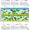
Dig-PCR of DNA from a stool sample. The 384 wells used in the experiment are displayed. Those colored blue contained 25 genome equivalents of DNA from normal cells. Each of these registered positive with MB-RED, and the RED/GREEN ratios were 1.0 ± 0.1 (mean ±1 SD). The wells colored yellow contained no template DNA, and each was negative with MB-RED (i.e., fluorescence <3,500 SFU.). The other 288 wells contained diluted DNA from the stool sample, prepared by alkaline extraction (57). Those registering positive with MB-RED were colored either red or green, depending on their RED/GREEN ratios. Those registering negative with MB-RED were colored white. PCR products from the indicated wells were used for automated sequence analysis.

Similar articles

Cited by

References

    1. Vogelstein B V, Kinzler K W. The Genetic Basis of Human Cancer. Toronto: McGraw–Hill; 1998.
    1. Lee C M, Weindruch R, Aiken J M. Free Radical Biol Med. 1997;22:1259–1269. - PubMed
    1. Ozawa T. Physiol Rev. 1997;77:425–464. - PubMed
    1. Sidransky D, Von Eschenbach A, Tsai Y C, Jones P, Summerhayes I, Marshall F, Paul M, Green P, Hamilton S R, Frost P, et al. Science. 1991;252:706–709. - PubMed
    1. Sidransky D, Tokino T, Hamilton S R, Kinzler K W, Levin B, Frost P, Vogelstein B. Science. 1992;256:102–105. - PubMed

Publication types