. 1999 Aug 3;96(16):9322-7.
doi: 10.1073/pnas.96.16.9322.

Influence of the high density lipoprotein receptor SR-BI on reproductive and cardiovascular pathophysiology

Affiliations

Influence of the high density lipoprotein receptor SR-BI on reproductive and cardiovascular pathophysiology

B Trigatti et al. Proc Natl Acad Sci U S A. .

Abstract

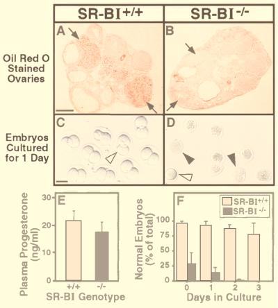
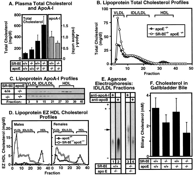
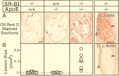
The high density lipoprotein (HDL) receptor SR-BI (scavenger receptor class B type I) mediates the selective uptake of plasma HDL cholesterol by the liver and steroidogenic tissues. As a consequence, SR-BI can influence plasma HDL cholesterol levels, HDL structure, biliary cholesterol concentrations, and the uptake, storage, and utilization of cholesterol by steroid hormone-producing cells. Here we used homozygous null SR-BI knockout mice to show that SR-BI is required for maintaining normal biliary cholesterol levels, oocyte development, and female fertility. We also used SR-BI/apolipoprotein E double homozygous knockout mice to show that SR-BI can protect against early-onset atherosclerosis. Although the mechanisms underlying the effects of SR-BI loss on reproduction and atherosclerosis have not been established, potential causes include changes in (i) plasma lipoprotein levels and/or structure, (ii) cholesterol flux into or out of peripheral tissues (ovary, aortic wall), and (iii) reverse cholesterol transport, as indicated by the significant reduction of gallbladder bile cholesterol levels in SR-BI and SR-BI/apolipoprotein E double knockout mice relative to controls. If SR-BI has similar activities in humans, it may become an attractive target for therapeutic intervention in a variety of diseases.


Figures

Figure 1
Figure 1
In vivo ovarian lipid accumulation in and in vitro development of preimplantation embryos from wild-type and SR-BI KO mice. Six-week-old female mice were superovulated and were mated to males of the other genotype (i.e., SR-BI+/+ females mated to SR-BI−/− males and vice versa) to generate embryos with heterozygous mutant genotypes. Ovaries and preimplantation embryos were harvested the next morning (day 0). (A and B) Typical oil red O staining of lipids in ovaries from SR-BI+/+ (A) or SR-BI−/− (B) animals. The arrows indicate corpora lutea. (Bar = 450 μm.) (C and D) Phase-contrast microscopy of preimplantation embryos (cultured for 1 day) from SR-BI+/+ (C) or SR-BI−/− (D) females mated to males of the opposite genotype. Similar results were observed when SR-BI−/− males were mated to SR-BI−/− females. Open arrowheads indicate morphologically normal, one- or two-cell embryos; solid arrowheads indicate embryos with abnormal, nonrefractive morphology. (Bar = 100 μm.) (E) Plasma progesterone concentrations from pseudopregnant females (6 days postmating, ages 6–10 weeks, weight = 19–25 g, n = 8; P = 0.08). (F) Percentage of preimplantation embryos from SR-BI+/+ (open bars) or SR-BI−/− (solid bars) females with normal morphology during 3 days of culture. The values represent the averages from five animals of each genotype. Total number of embryos: SR-BI+/+, 131; SR-BI−/−, 167.
Figure 2
Figure 2
Effects of SR-BI gene disruption on plasma lipoproteins in apoE KO mice. Mice were 4–7 weeks old. (A) Plasma total cholesterol levels (Left, mean ± SD) for wild-type (n = 5), SR-BI−/− (n = 12), apoE−/− (n = 12), and SR-BI−/− apoE−/− (n = 9) females (P < 0.0002). Plasma apoA-I levels (Right, mean ± SD, expressed as relative units) were determined by SDS/PAGE (15%) followed by quantitative immunoblotting for apoE−/− (n = 7) and SR-BI−/−apoE−/− females (n = 5) (P = 0.1). (B) Lipoprotein cholesterol profiles. Plasma lipoproteins from individual apoE−/− (shaded diamonds) or SR-BI−/− apoE−/− (○) females were separated based on size (Superose 6-FPLC), and total cholesterol in each fraction (expressed as mg/dl plasma) was measured. The chromatograms shown are representative of multiple, independent determinations. Approximate elution positions of VLDL, IDL/LDL, and HDL are indicated as described previously (17, 18). (C) Lipoprotein apoA-I profiles. Pooled Superose 6-FPLC fractions (see above, ≈21 μl per pool) from females in an independent experiment were analyzed by SDS-polyacrylamide gradient (3–15%) gel electrophoresis and immunoblotting with an anti-apoA-I antibody (18). Each pool contained three fractions, and lanes are labeled with the number of the middle fraction in each pool. (D) Average EZ HDL cholesterol FPLC profiles for apoE−/− (diamonds) or SR-BI−/− apoE−/− (○) males (Left, n = 3) or females (Right, n = 3). (E) Agarose gel electrophoresis and immunoblotting. Pooled fractions (fractions 11–21; 3.5 μl) from the IDL/LDL region of the lipoprotein profile from individual apoE−/− or SR-BI−/− apoE−/− females were analyzed by using either anti-apoA-I or anti-apoB antibodies. The position of migration of normal-sized HDL is indicated by the asterisk. The arrow on the left indicates the mobility of the apoA-I-containing, apoB-free particles from double KOs (lane 3). Migration was upward from negative to positive (origin not shown). (F) Gallbladder biliary cholesterol (mean ± SD). Total gallbladder biliary cholesterol from both male and female mice of the indicated genotypes (n = 10 or 11 per genotype) was measured. Except for the wild-type and apoE−/− values, all pairwise differences were statistically significant (P < 0.025–0.0005).
Figure 3
Figure 3
Effects of SR-BI gene disruption on atherosclerosis in apoE KO mice. Atherosclerosis in SR-BI−/− (n = 8, 4–6 weeks old), apoE−/− (n = 8, 5–7 weeks old), or SR-BI−/− apoE−/− (n = 7, 5–6 weeks old) female mice was analyzed in cryosections of aortic sinuses stained with oil red O and Mayer’s hematoxylin as described in Materials and Methods. (A) Representative sections through the aortic root region. (Bar = 200 μm.) (B) Sizes (cross-sectional areas) of oil red O-stained lesions in the aortic root region (see Materials and Methods). Average lesion areas (mm2 ± SD) for SR-BI−/−apoE−/−, apoE−/−, or SR-BI−/− mice, respectively, were as follows: 0.10 ± 0.07 (horizontal line), 0.002 ± 0.002, and 0.001 ± 0.002 (P = 0.0005). Also see Table 1. (C and D) High-magnification views of serial sections of plaque from the aortic sinus of a 7-week-old SR-BI/apoE double KO male, stained either with oil red O and Mayer’s hematoxylin (C) or with an anti-α actin antibody that recognizes smooth-muscle cells (D). The lumen is to the left of the plaque. The smooth-muscle wall (arrowheads) and cellular cap (arrows) are indicated. (Bar = 100 μm.)

References

    1. Gordon D J, Rifkind B M. N Engl J Med. 1989;321:1311–1316. - PubMed
    1. Johnson W J, Mahlberg F H, Rothblat G H, Phillips M C. Biochim Biophys Acta. 1991;1085:273–298. - PubMed
    1. Tall A R. J Lipid Res. 1993;34:1255–1274. - PubMed
    1. Pieters M N, Schouten D, VanBerkel T J C. Biochim Biophys Acta. 1994;1225:125–134. - PubMed
    1. Fielding C J, Fielding P E. J Lipid Res. 1995;36:211–228. - PubMed

Publication types

MeSH terms