. 1999 Aug 3;96(16):9345-50.
doi: 10.1073/pnas.96.16.9345.

Generation of influenza A viruses entirely from cloned cDNAs

Affiliations

Generation of influenza A viruses entirely from cloned cDNAs

G Neumann et al. Proc Natl Acad Sci U S A. .

Abstract

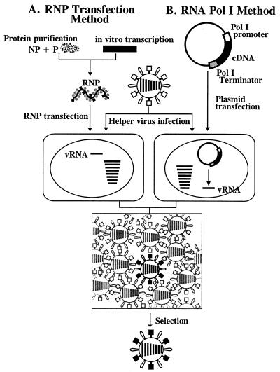
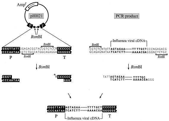
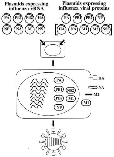
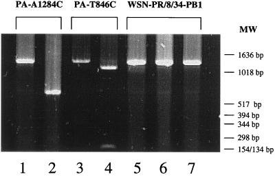
We describe a new reverse-genetics system that allows one to efficiently generate influenza A viruses entirely from cloned cDNAs. Human embryonic kidney cells (293T) were transfected with eight plasmids, each encoding a viral RNA of the A/WSN/33 (H1N1) or A/PR/8/34 (H1N1) virus, flanked by the human RNA polymerase I promoter and the mouse RNA polymerase I terminator-together with plasmids encoding viral nucleoprotein and the PB2, PB1, and PA viral polymerases. This strategy yielded >1 x 10(3) plaque-forming units (pfu) of virus per ml of supernatant at 48 hr posttransfection. The addition of plasmids expressing all of the remaining viral structural proteins led to a substantial increase in virus production, 3 x 10(4)-5 x 10(7) pfu/ml. We also used reverse genetics to generate a reassortant virus containing the PB1 gene of the A/PR/8/34 virus, with all other genes representing A/WSN/33. Additional viruses produced by this method had mutations in the PA gene or possessed a foreign epitope in the head of the neuraminidase protein. This efficient system, which does not require helper virus infection, should be useful in viral mutagenesis studies and in the production of vaccines and gene therapy vectors.


Figures

Figure 1
Figure 1
Schematic diagram of established reverse-genetics systems. In the RNP transfection method (A), purified NP and polymerase proteins are assembled into RNPs with the use of in vitro-synthesized vRNA. Cells are transfected with RNPs, followed by helper virus infection. In the RNA polymerase I method (B), a plasmid containing the RNA polymerase I promoter, a cDNA encoding the vRNA to be rescued, and the RNA polymerase I terminator are transfected into cells. Intracellular transcription by RNA polymerase I yields synthetic vRNA, which is packaged into progeny virus particles upon infection with helper virus. With both methods, transfectant viruses (i.e., those containing RNA derived from cloned cDNA) are selected from the helper virus population.
Figure 2
Figure 2
Schematic diagram of the generation of RNA polymerase I constructs. cDNAs derived from influenza virus were amplified by PCR, digested with BsmBI, and cloned into the BsmBI sites of the pHH21 vector (34), which contains the human RNA polymerase I promoter (P) and the mouse RNA polymerase I terminator (T). The thymidine nucleotide upstream of the terminator sequence (*T) represents the 3′ end of the influenza viral RNA. Influenza A virus sequences are shown in boldface letters.
Figure 3
Figure 3
Reverse-genetics method for generating segmented, negative-sense RNA viruses entirely from cloned cDNA. Plasmids containing the RNA polymerase I promoter, a cDNA for each of the eight viral RNA segments, and the RNA polymerase I terminator are transfected into cells together with protein expression plasmids. Although infectious viruses can be generated with plasmids expressing PA, PB1, PB2, and NP, expression of all remaining structural proteins (shown in brackets) increases the efficiency of virus production (see text).
Figure 4
Figure 4
Detection of the Flag epitope in cells infected with a transfectant virus. Antibody staining was used to identify the NA in MDCK cells infected with either PR8-WSN-FL79 (A and D) or A/WSN/33 wild-type virus (B and E) or on mock-infected MDCK cells (C and F). Nine hours after infection, cells were fixed with paraformaldehyde, treated with Triton X-100, and incubated with either anti-Flag (AC) or anti-WSN NA (DF) mAbs. Intensive Golgi staining (red) is apparent in positive samples (A, D, and E).
Figure 5
Figure 5
Recovery of PA mutants. The PA gene of each virus was amplified by reverse transcriptase–PCR with primers that yield a 1,226-bp fragment (position 677-1903 of the mRNA; lanes 1, 3, and 5), which then was digested with the restriction enzyme Bsp120I (at position 846 of the mRNA; lanes 4 and 7) or PvuII (at position 1284 of the mRNA; lanes 2 and 6). The presence of Bsp120I or PvuII sites in the PCR products yielded either 169- and 1,057-bp or 607- and 619-bp fragments, respectively. MW, molecular weight markers.

Comment in

References

    1. Palese P, Zheng H, Engelhardt O G, Pleschka S, Garcia-Sastre A. Proc Natl Acad Sci USA. 1996;93:11354–11358. - PMC - PubMed
    1. Schnell M J, Mebatsion T, Conzelmann K K. EMBO J. 1994;13:4195–4203. - PMC - PubMed
    1. Lawson N D, Stillman E A, Whitt M A, Rose J K. Proc Natl Acad Sci USA. 1995;92:4477–4481. - PMC - PubMed
    1. Whelan S P, Ball L A, Barr J N, Wertz G T. Proc Natl Acad Sci USA. 1995;92:8388–8392. - PMC - PubMed
    1. Radecke F, Spielhofer P, Schneider H, Kaelin K, Huber M, Dotsch C, Christiansen G, Billeter M A. EMBO J. 1995;14:5773–5784. - PMC - PubMed

Publication types