. 1999 Aug 3;96(16):9379-84.
doi: 10.1073/pnas.96.16.9379.

Distributed representation of objects in the human ventral visual pathway

Affiliations

Distributed representation of objects in the human ventral visual pathway

A Ishai et al. Proc Natl Acad Sci U S A. .

Abstract

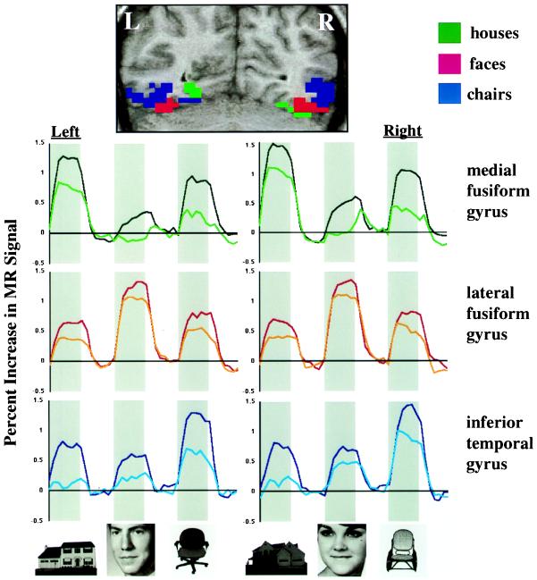
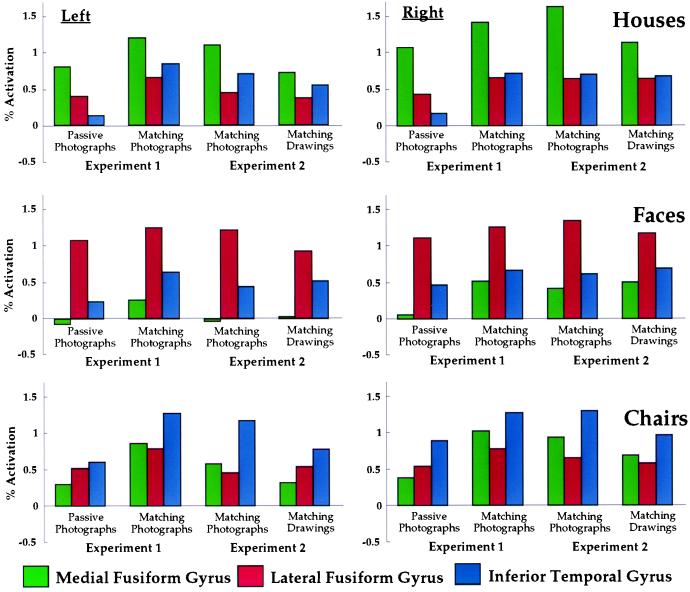
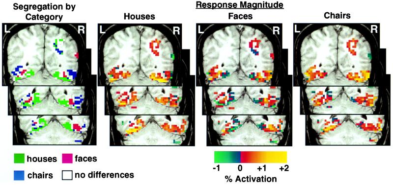
Brain imaging and electrophysiological recording studies in humans have reported discrete cortical regions in posterior ventral temporal cortex that respond preferentially to faces, buildings, and letters. These findings suggest a category-specific anatomically segregated modular organization of the object vision pathway. Here we present data from a functional MRI study in which we found three distinct regions of ventral temporal cortex that responded preferentially to faces and two categories of other objects, namely houses and chairs, and had a highly consistent topological arrangement. Although the data could be interpreted as evidence for separate modules, we found that each category also evoked significant responses in the regions that responded maximally to other stimuli. Moreover, each category was associated with its own differential pattern of response across ventral temporal cortex. These results indicate that the representation of an object is not restricted to a region that responds maximally to that object, but rather is distributed across a broader expanse of cortex. We propose that the functional architecture of the ventral visual pathway is not a mosaic of category-specific modules but instead is a continuous representation of information about object form that has a highly consistent and orderly topological arrangement.


Figures

Figure 1
Figure 1
Response topographies in ventral temporal cortex in Experiment 1. (Top) Locations of three posterior ventral temporal regions that responded differentially to houses, faces, and chairs, illustrated in a coronal section (y = −65) from a single subject. Voxels shown in color demonstrated a significant overall experimental effect (Z > 4.0) and a significant difference among responses to houses, faces, and chairs (Z > 1.96, clusters of seven or more voxels). Regions showing maximal responses to houses, faces, and chairs are shown in green, red, and blue, respectively. (Bottom) Mean time series for these three ventral temporal regions. Data are averaged across six subjects and 12 repetitions of task blocks in each subject. Gray bars indicate presentation of meaningful stimuli. The white space to the right of each gray bar indicates the presentation of control stimuli. The darker colored line in each graph is for the delayed match-to-sample task, and the lighter line is for the passive viewing task.
Figure 2
Figure 2
Patterns of response to houses, faces, and chairs across three regions in posterior ventral temporal cortex. Results from Experiments 1 and 2 are shown in the same graphs to allow direct comparison of the patterns of response.
Figure 3
Figure 3
Patterns of response to houses, faces, and chairs in three coronal sections through ventral temporal cortex in one subject. Regions showing maximal responses to houses, faces, and chairs, are shown in green, red, and blue, respectively. White voxels indicate significant activation across stimulus categories but no significant differences between categories. Strength of response to houses, faces, and chairs relative to control tasks with scrambled pictures is shown in all ventral temporal voxels that showed a significant experimental effect (Z > 4.0). (Top) y = −60; (Middle) y = −55; (Bottom) y = −50.

References

    1. Allison T, McCarthy G, Nobre A, Puce A, Belger A. Cereb Cortex. 1994;5:544–554. - PubMed
    1. Puce A, Allison T, Gore J C, McCarthy G. J Neurophysiol. 1995;74:1192–1199. - PubMed
    1. Puce A, Allison T, Asgari M, Gore J C, McCarthy G. J Neurosci. 1996;16:5205–5215. - PMC - PubMed
    1. Clark V P, Keil K, Maisog J M, Courtney S, Ungerleider L G, Haxby J V. Neuroimage. 1996;4:1–15. - PubMed
    1. Kanwisher N, McDermott J, Chun M M. J Neurosci. 1997;17:4302–4311. - PMC - PubMed

LinkOut - more resources