. 1999 Aug 3;96(16):9385-90.
doi: 10.1073/pnas.96.16.9385.

Neurotrophins: peripherally and centrally acting modulators of tactile stimulus-induced inflammatory pain hypersensitivity

Affiliations

Neurotrophins: peripherally and centrally acting modulators of tactile stimulus-induced inflammatory pain hypersensitivity

R J Mannion et al. Proc Natl Acad Sci U S A. .

Abstract

Brain-derived neurotrophic factor (BDNF) is expressed in nociceptive sensory neurons and transported anterogradely to the dorsal horn of the spinal cord where it is located in dense core vesicles in C-fiber terminals. Peripheral inflammation substantially up-regulates BDNF mRNA and protein in the dorsal root ganglion (DRG) in a nerve growth factor-dependent fashion and results in novel expression of BDNF by DRG neurons with myelinated axons. C-fiber electrical activity also increases BDNF expression in the DRG, and both inflammation and activity increase full-length TrkB receptor levels in the dorsal horn. Sequestration of endogenous BDNF/neurotrophin 4 by intraspinal TrkB-Fc fusion protein administration does not, in noninflamed animals, change basal pain sensitivity nor the mechanical hypersensitivity induced by peripheral capsaicin administration, a measure of C fiber-mediated central sensitization. TrkB-Fc administration also does not modify basal inflammatory pain hypersensitivity, but does block the progressive hypersensitivity elicited by low-intensity tactile stimulation of inflamed tissue. BDNF, by virtue of its nerve growth factor regulation in sensory neurons including novel expression in A fibers, has a role as a central modulator of tactile stimulus-induced inflammatory pain hypersensitivity.


Figures
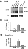
Figure 1
Figure 1
Change in BDNF mRNA and protein levels in L4 and L5 DRGs and lumbar dorsal horn. (A) Northern blots for BDNF and cyclophilin mRNA in L4 and L5 DRGs showing increased BDNF mRNA after stimulation of the sciatic nerve at C- but not A-fiber strength and 24 h after inflammation of the hindpaw. (B) BDNF protein levels, measured by ELISA, in L4 and L5 DRGs (n = 6) in naïve and inflamed rats (48 h post-CFA). Inflammation increased BDNF in the ipsilateral DRG (P < 0.01), which was prevented by anti-NGF (P < 0.01). (C) The elevation of BDNF in the dorsal horn 48 h after inflammation (n = 6, P < 0.05) also was prevented by anti-NGF pretreatment (n = 6, P < 0.05).
Figure 2
Figure 2
Increase and phenotypic change in BDNF expression in the DRG. BDNF mRNA labeled neuronal profiles increase 2 h after stimulation of the sciatic nerves (B) and 24 h after inflammation (C). The number of labeled profiles/total neuronal profiles per section increased from 11.1 ± 1.7 (naives, A) to 31.4 ± 2.4 (C-fiber stimulation, B) and 35.9 ± 9.2 (inflammation, C) (n = 3, P < 0.05). Scale (A–C): 100 μm. Profile area-frequency histograms constructed from 600 profiles in three animals show an increase in the number of small and medium sized neuronal profiles expressing BDNF mRNA. Cells >600 μm2 (shaded) are A fibers (29). (D) Double staining for BDNF mRNA and NF200 immunohistochemistry shows only a small proportion of BDNF mRNA-expressing cells are NF200 positive in naives (arrows), but after inflammation the proportion of double-labeled cells increases 2-fold. Scale (D): 200 μm.
Figure 3
Figure 3
TrkB mRNA and protein up-regulation in the dorsal horn. (A) Northern blots for full-length TrkB mRNA show up-regulation of this transcript after inflammation (CFA). Electrical stimulation (Stim) also induces up-regulation of full-length TrkB mRNA. Cyc, cyclophilin. (B) Western blot for TrkB protein shows up-regulation of both full-length and truncated TrkB in the dorsal horn at 3 and 5 days post-CFA. (C) In situ hybridization for full-length TrkB mRNA in the dorsal horn shows labeling in all laminae. Scale: 200 μm.
Figure 4
Figure 4
Intrathecal TrkB-Fc, but not TrkC-Fc, fusion protein prevents the establishment of progressive tactile hypersensitivity. (A and B) Electrophysiological recordings from single flexor motoneurons showing that in TrkC-Fc-, but not TrkB-Fc-, treated animals tactile stimulation of the inflamed paw progressively reduces mechanical threshold and touch responsiveness (n = 6, P < 0.001). In behaving animals TkB-Fc attenuates the reduction in mechanical threshold for flexion withdrawal during the period of intermittent tactile stimulation (n = 7) compared with TrkC-Fc-treated animals (n = 7) (C) A cumulative response plot of the thresholds in the two treatment groups 20 min after commencement of the tactile stimuli shows a significant shift of the curve to the left (P = 0.005) (D).

References

    1. Apfel S C, Wright D E, Wiideman A M, Dormia C, Snider W D, Kessler J A. Mol Cell Neurosci. 1996;7:134–142. - PubMed
    1. Yan Q, Rosenfeld R D, Matheson C R, Lopez O T, Bennett L, Welcher A A. Neuroscience. 1997;78:431–448. - PubMed
    1. Zhou X F, Rush R A. Neuroscience. 1996;74:945–953. - PubMed
    1. Michael G J, Averill S, Nitkunan A, Rattray M, Bennett D L H, Yan Q, Priestley J V. J Neurosci. 1997;17:8476–8490. - PMC - PubMed
    1. Korte M, Carroll P, Wolf E, Brem G, Thoenen H, Bonhoeffer T. Proc Natl Acad Sci USA. 1995;92:8856–8860. - PMC - PubMed

Publication types

MeSH terms

Substances