Case Reports
. 1999 Jul 19;190(2):183-94.
doi: 10.1084/jem.190.2.183.

A novel H(+) conductance in eosinophils: unique characteristics and absence in chronic granulomatous disease

Affiliations
Case Reports

A novel H(+) conductance in eosinophils: unique characteristics and absence in chronic granulomatous disease

B Bánfi et al. J Exp Med. .

Abstract

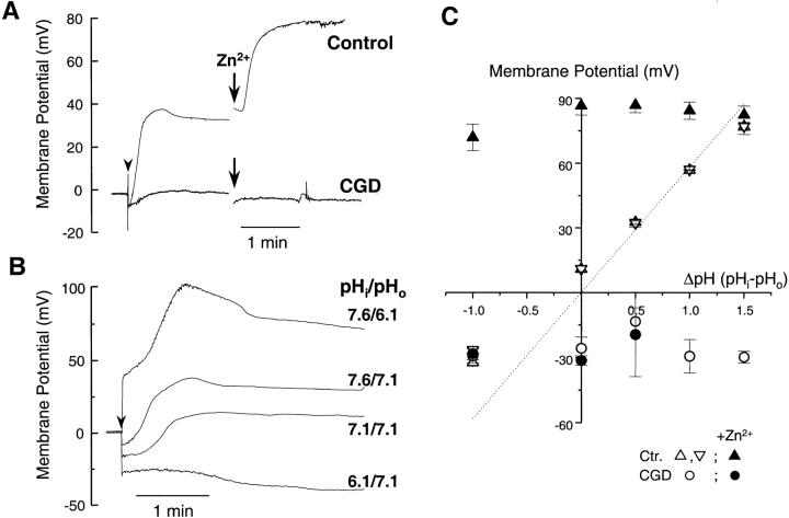
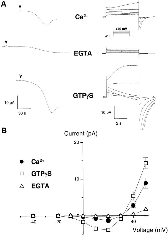
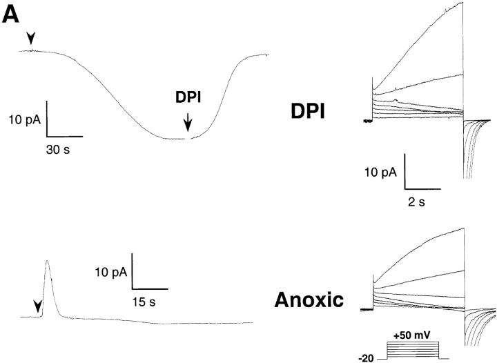
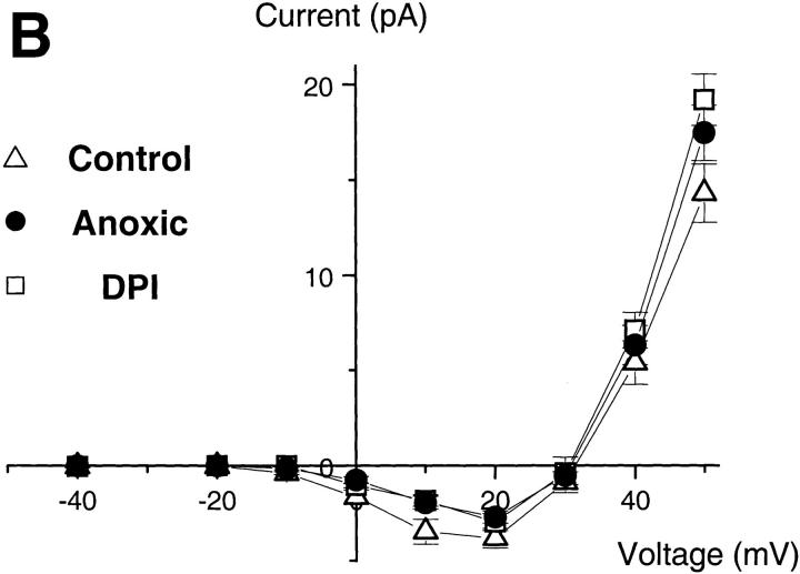
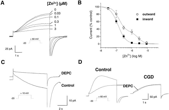
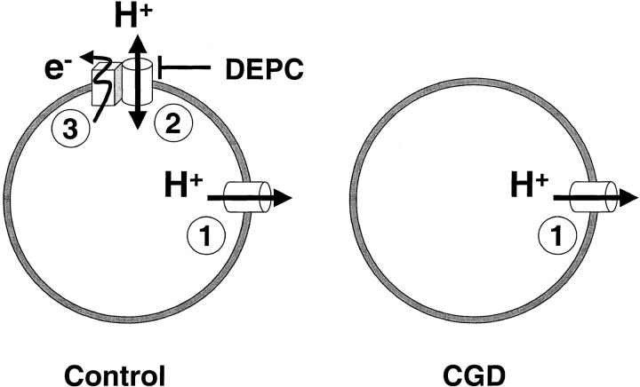
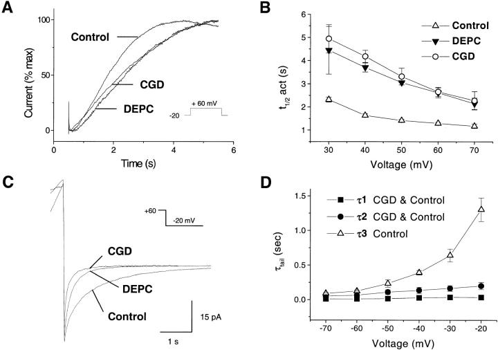
Efficient mechanisms of H(+) ion extrusion are crucial for normal NADPH oxidase function. However, whether the NADPH oxidase-in analogy with mitochondrial cytochromes-has an inherent H(+) channel activity remains uncertain: electrophysiological studies did not find altered H(+) currents in cells from patients with chronic granulomatous disease (CGD), challenging earlier reports in intact cells. In this study, we describe the presence of two different types of H(+) currents in human eosinophils. The "classical" H(+) current had properties similar to previously described H(+) conductances and was present in CGD cells. In contrast, the "novel" type of H(+) current had not been described previously and displayed unique properties: (a) it was absent in cells from gp91- or p47-deficient CGD patients; (b) it was only observed under experimental conditions that allowed NADPH oxidase activation; (c) because of its low threshold of voltage activation, it allowed proton influx and cytosolic acidification; (d) it activated faster and deactivated with slower and distinct kinetics than the classical H(+) currents; and (e) it was approximately 20-fold more sensitive to Zn(2+) and was blocked by the histidine-reactive agent, diethylpyrocarbonate (DEPC). In summary, our results demonstrate that the NADPH oxidase or a closely associated protein provides a novel type of H(+) conductance during phagocyte activation. The unique properties of this conductance suggest that its physiological function is not restricted to H(+) extrusion and repolarization, but might include depolarization, pH-dependent signal termination, and determination of the phagosomal pH set point.


Figures

Figure 1
Figure 1
Membrane potential changes during activation of control and CGD eosinophils. The membrane potential of eosinophils, determined as the zero current holding potential, was measured in current clamp mode after the formation of the whole cell configuration (arrowhead). The pipette solutions contained 8 mM NADPH and 25 μM GTPγS to activate the oxidase. (A) Time course of the membrane potential changes in control and CGD eosinophils, measured in Cs+-based solutions containing 10 mM TEA (pHi = 7.6, pHo = 7.1). When indicated, the H+ conductance blocker, Zn2+ (10 μM), was added to the bath solution. (B) Membrane potential changes measured with CsCl solutions buffered to different pH. (C) The steady state membrane potential of control (triangles) and CGD cells (circles) is plotted against the transmembrane pH gradient, measured in CsCl (upward triangles) or quasiphysiological (downward triangles) solutions, in the absence (open symbols) or presence (filled symbols) of Zn2+. The dotted line represents the calculated H+ equilibrium potential. Data are mean ± SEM of ≥5 experiments for each condition.
Figure 2
Figure 2
H+ currents during activation of the NADPH oxidase. (A) Whole cell current in human eosinophils measured under resting and stimulated conditions. The pipette solution contained 8 mM NADPH to provide substrate for the oxidase, and either no calcium buffer (top, ∼5 μM free [Ca2+]), 10 mM EGTA (middle), or 25 μM GTPγS (bottom). After break-in (arrowheads), the electron currents were recorded at 0 mV (left traces). After achieving a steady state, the holding voltage was changed to –20 mV, and 5-s depolarizing steps ranging from –10 to +40 mV (inset) were applied in 10-mV increments to elicit H+ currents (right traces). Traces are representative of ≥20 experiments for each condition. (B) Current–voltage plot of the voltage-dependent currents measured with ∼5 μM free [Ca2+] (•), 25 μM GTPγS (□), and 10 mM EGTA (▵). Currents measured 500 ms after the beginning of a 5-s-long voltage pulse were subtracted from the current measured at the end of the pulse. The leak-subtracted currents reversed sign around +30 mV, close to the H+ reversal potential (pHi = 7.6, pHo = 7.1; EH+ = +29 mV; mean ± SEM of ≥7 experiments).
Figure 5
Figure 5
The inward H+ currents do not require oxidase activity. (A) Effect of DPI (top) and of oxygen depletion (bottom) on the electron (left) and proton currents (right traces). Pipette solutions contained 25 μM GTPγS, 8 mM NADPH, pH 7.6; bath pH 7.1. Oxygen was removed from the bath solution by a 4-h preincubation with glucose-oxidase (50 mU/ml) and catalase (2,000 U/ml). Traces are representative of ≥7 experiments. (B) Current–voltage relationship of the H+ currents, measured as in the legend to Fig. 2, under control conditions (▵, PO2 = 25.7 ± 0.3 kPa), in the presence of DPI (□), or under low oxygen conditions (•, PO2 < 1 kPa). Data are mean ± SEM of ≥7 experiments for each condition.
Figure 5
Figure 5
The inward H+ currents do not require oxidase activity. (A) Effect of DPI (top) and of oxygen depletion (bottom) on the electron (left) and proton currents (right traces). Pipette solutions contained 25 μM GTPγS, 8 mM NADPH, pH 7.6; bath pH 7.1. Oxygen was removed from the bath solution by a 4-h preincubation with glucose-oxidase (50 mU/ml) and catalase (2,000 U/ml). Traces are representative of ≥7 experiments. (B) Current–voltage relationship of the H+ currents, measured as in the legend to Fig. 2, under control conditions (▵, PO2 = 25.7 ± 0.3 kPa), in the presence of DPI (□), or under low oxygen conditions (•, PO2 < 1 kPa). Data are mean ± SEM of ≥7 experiments for each condition.
Figure 3
Figure 3
H+ current–associated changes in cytosolic pH. Combined recordings of whole cell currents and cytosolic pH changes measured with the fluorescent pH indicator carboxy-SNARF-1. The pipette solutions contained 25 μM GTPγS and 8 mM NADPH. (A) After break-in (arrowhead), the cytosol was allowed to equilibrate with the alkaline pipette solution (pH = 7.6). Then, long-lasting (20 s) depolarizing steps to +20 mV were applied (middle), and the currents (bottom) and cytosolic pH changes (top) were measured concomitantly. When indicated, Zn2+ (10 μM) was added to the bath solution. (B) A sustained depolarization to +80 mV was imposed (middle) to allow H+ efflux through the conductance (pHi = 8.1, pHo = 7.1, EH+ = +58 mV). After establishing a new steady state pH (top) and current (bottom), the external pH was rapidly decreased from 7.1 to 6.6 to change the direction of the H+ gradient (EH+ = +90 mV). Recordings are representative of ≥16 experiments for each condition.
Figure 4
Figure 4
pH dependence of the H+ currents. (A) Currents elicited by a 5-s depolarizing pulse to +10 mV in cells perfused with alkaline (pH = 7.6, n = 20) or neutral (pH = 7.1, n = 7) pipette solutions containing 25 μM GTPγS. (B) Current–voltage relationship measured at a pipette pH of 6.1 (▾), 7.1 (▵), 7.6 (•), and 8.1 (□). The holding voltage was –60 mV (pH 6.1), −40 mV (pH 7.1), and –20 mV (pH 7.6 and 8.1), and bath pH was 7.1 in all conditions. Arrows indicate the H+ equilibrium potential for each condition. Data are mean ± SEM of ≥7 experiments for each condition.
Figure 4
Figure 4
pH dependence of the H+ currents. (A) Currents elicited by a 5-s depolarizing pulse to +10 mV in cells perfused with alkaline (pH = 7.6, n = 20) or neutral (pH = 7.1, n = 7) pipette solutions containing 25 μM GTPγS. (B) Current–voltage relationship measured at a pipette pH of 6.1 (▾), 7.1 (▵), 7.6 (•), and 8.1 (□). The holding voltage was –60 mV (pH 6.1), −40 mV (pH 7.1), and –20 mV (pH 7.6 and 8.1), and bath pH was 7.1 in all conditions. Arrows indicate the H+ equilibrium potential for each condition. Data are mean ± SEM of ≥7 experiments for each condition.
Figure 7
Figure 7
Block of the currents by Zn2+ and DEPC. Effect of the polyvalent cation Zn2+ and of the histidine reagent DEPC on the H+ currents of control and CGD cells. Pipette solutions contained 25 μM GTPγS, pHi 7.6 (A–C) or 7.1 (D), pHo 7.1. (A) Effect of Zn2+ on the outward currents in control, activated eosinophils. Currents were elicited by a pulse from −20 to +60 mV, and increasing free concentrations of Zn2+ (buffered with 5 mM citrate) were added to the bath solution. Traces are representative of ≥11 experiments. (B) Dose-inhibition curves of the block by Zn2+. The fractional outward (○, measured at +60 mV) or inward (▪, measured at +20 mV) current is plotted against the extracellular Zn2+ concentration. Data are mean ± SEM of ≥4 experiments for each condition, fitted with a single (▪) or double sigmoidal curve (○) using Origin software. (C) Effect of DEPC (1.2 mM) on the inward current induced by a 7-s-long depolarization to +10 mV. (D) Effect of DEPC on the outward current induced by a 5-s depolarization to +60 mV. Traces are representative of ≥7 experiments.
Figure 6
Figure 6
The inward H+ currents are absent in CGD patients. Proton currents recorded in eosinophils from two CGD patients: a 6-yr-old Caucasian boy with X-linked CGD, who completely lacked the gp91phox subunit (X910), and a 34-yr-old Caucasian woman with a deficiency in p47 (A470). (A) Families of currents elicited by 5-s depolarizing steps ranging from 0 mV (CGD) or −20 mV (Control) to +50 mV, in conditions favoring inward currents (pHi = 7.6, 25 μM GTPγS). (B) Currents elicited by 5-s pulses from −60 mV to 0 mV, in conditions minimizing inward currents (pHi 6.1, 0.2 mM EGTA, no GTPγS). Traces are representative of ≥8 experiments. (C) Current–voltage plot of proton currents measured in CGD cells under different activating conditions (pHi = 7.6, pHo = 7.1). Data are mean ± SEM of ≥4 experiments for each condition.
Figure 9
Figure 9
CGD eosinophils lack a distinct H+ conductance linked to the oxidase. Diagram illustrating the two types of H+ conductances described in this study, and their coupling to the NADPH oxidase. The “classical” H+ conductance 1 is present in both control and CGD cells and allows only H+ extrusion and repolarization. This conductance activates slowly, deactivates rapidly, is blocked by Zn2+ with low affinity, and is insensitive to DEPC. In contrast, the novel H+ conductance 2 is absent in CGD cells and allows H+ influx and depolarization. It activates rapidly, inactivates slowly, is highly sensitive to Zn2+, and is blocked by DEPC. This conductance is closely coupled to and might possibly be part of the NADPH oxidase 3.
Figure 8
Figure 8
Activation and deactivation of the currents in control and CGD cells. pHi 7.1 (A, B) or 7.6 (C, D), pHo 7.1. (A) Kinetics of current activation in control, CGD, and DEPC-treated cells. Currents elicited by a 5-s-long pulse from –20 mV to +60 mV were normalized to the maximal current recorded at this voltage and superimposed for comparison. (B) Voltage dependence of current activation. The time for half-maximal activation (t 1/2 act) is plotted against the activating voltage. (C) Kinetics of current deactivation during repolarization from +60 mV to –20 mV. Cells were depolarized for various durations to induce currents of similar amplitude. Traces are representative of ≥9 experiments. (D) Voltage dependence of the deactivation time constants (τtail), estimated by fitting exponential curves to the currents measured after a pulse to +60 mV. The two fast kinetic components (τ1, ▪; τ2, •) are present in both control and CGD cells, whereas an additional third slow component (τ3, ▵) is absent from CGD cells. Data are mean ± SEM of ≥5 experiments for each condition.

References

    1. Babior B.M. Oxygen-dependent microbial killing by phagocytes (first of two parts) N. Engl. J. Med. 1978;298:659–668. - PubMed
    1. Klebanoff S.J. Oxygen metabolism and the toxic properties of phagocytes. Ann. Intern. Med. 1980;93:480–489. - PubMed
    1. Segal A.W. The antimicrobial role of the neutrophil leukocyte. J. Infect. 1981;3:3–17. - PubMed
    1. Clark R.A. The human neutrophil respiratory burst oxidase. J. Infect. Dis. 1990;161:1140–1147. - PubMed
    1. Segal A.W., Abo A. The biochemical basis of the NADPH oxidase of phagocytes. Trends Biochem. Sci. 1993;18:43–47. - PubMed

Publication types

MeSH terms