Comparative Study
. 1999 Aug 15;212(2):323-36.
doi: 10.1006/dbio.1999.9335.

Vertebrate homologs of Drosophila suppressor of fused interact with the gli family of transcriptional regulators

Affiliations
Comparative Study

Vertebrate homologs of Drosophila suppressor of fused interact with the gli family of transcriptional regulators

R V Pearse 2nd et al. Dev Biol. .

Abstract

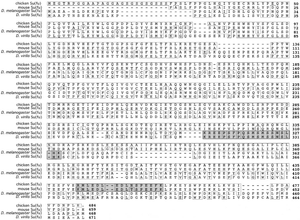
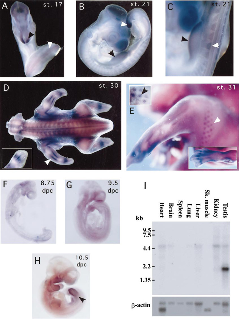
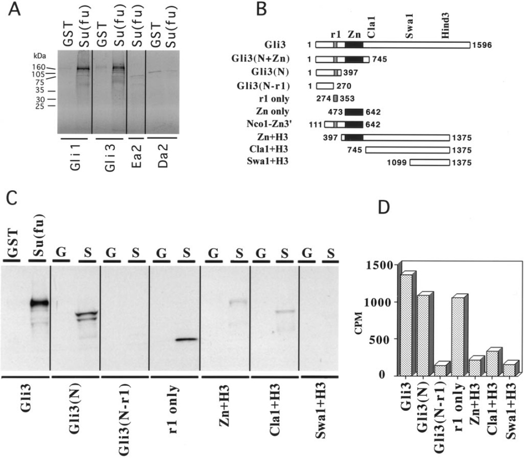
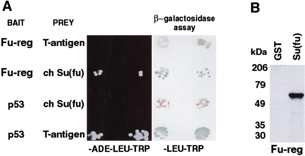
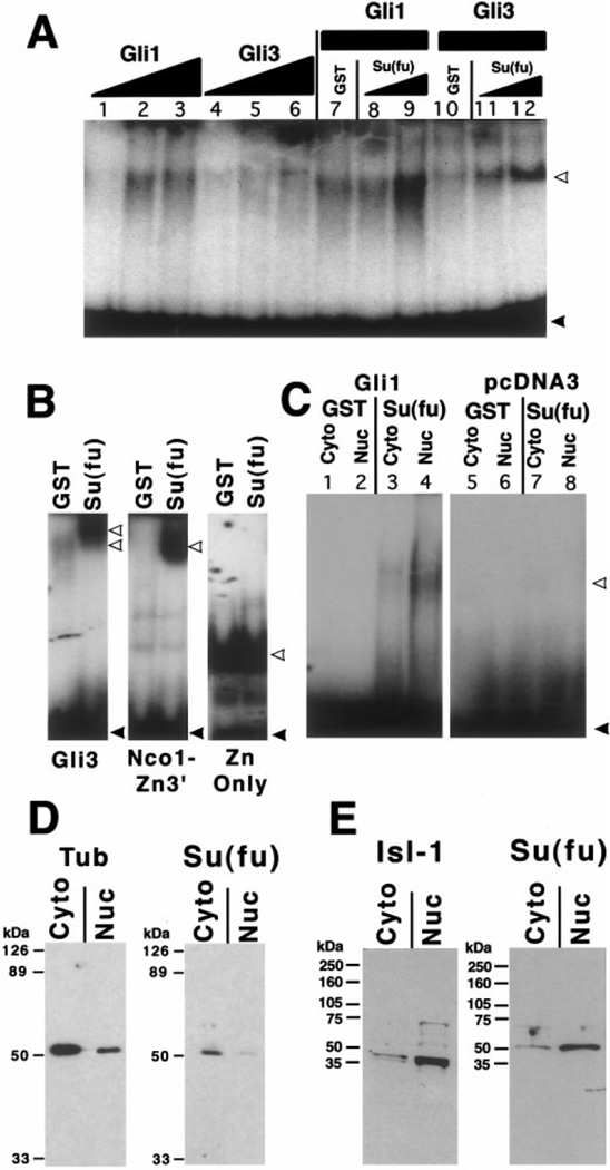
The hedgehog (Hh) signaling pathway is crucial for pattern formation during metazoan development. Although originially characterized in Drosophila, vertebrate homologs have been identified for several, but not all, genes in the pathway. Analysis of mutants in Drosophila demonstrates that Suppressor of fused [Su(fu)] interacts genetically with genes encoding proteins in the Hh signal transduction pathway, and its protein product physically interacts with two of the proteins in the Hh pathway. We report here the molecular cloning and characterization of chicken and mouse homologs of Su(fu). The chick and mouse proteins are 27% identical and 53% similar at the amino acid level to the Drosophila melanogaster and Drosophila virilis proteins. Vertebrate Su(fu) is widely expressed in the developing embryo with higher levels in tissues that are known to be patterned by Hh signaling. The chick Su(fu) protein can physically interact with factors known to function in Hh signal transduction including the Drosophila serine/threonine kinase, Fused, and the vertebrate transcriptional regulators Gli1 and Gli3. This interaction may be significant for transcriptional regulation, as recombinant Su(fu) enhances the ability of Gli proteins to bind DNA in electrophoretic mobility shift assays.


Figures

FIG. 1
FIG. 1
Sequence comparison between Drosophila and vertebrate Su(fu) proteins. Chicken and mouse amino acid sequences were aligned with the published Su(fu) sequences for D. melanogaster and D. virilis. Amino acids with identity to chick Su(fu) are boxed. Chick Su(fu) sequence extends from the initial methionine to the stop codon and contains an additional 15 amino acids at the amino-terminus compared to the Drosophila proteins. Numbers on the right indicate the position of the last amino acid in that row with the exception of mouse Su(fu), where we have not identified the initial methionine. For mouse Su(fu) position 1 is the most amino-terminal amino acid. The first shaded box illustrates the previously identified PEST sequences in Drosophila, and the second shaded box illustrates the amino acid sequences in chick and mouse Su(fu) that scored +1.51 and +4.07, respectively, using the PESTfind algorithm (http://www.at.embnet.org/htbin/embnet/PESTfind). The PEST sequences in Drosophila share no similarity to those in the vertebrate proteins.
FIG. 2
FIG. 2
Expression patterns of chick and mouse Su(fu). Su(fu) expression in the chick (A, B, C, D, E) and in mouse (F, G, H) embryos and in mouse adult tissues (I). (A) Hamburger–Hamilton stage 17 chick embryo showing Su(fu) expression at low levels in the neural tube (white arrow) with higher expression in the anterior tip of the neural epithelium (black arrow). (B) Stage 21 embryo showing slightly higher Su(fu) expression in the developing limb buds (black arrow) and in the tail bud (white arrow). (C) Higher magnification of limb bud (black arrow) and somite (white arrow) expression in stage 21 embryos. (D) Stage 30 chick embryo showing high levels of Su(fu) expression in the regions of the developing perichondrium. White arrow shows staining in the developing stylopod (forearm) in the region of the perichondrium and inset shows staining in the developing autopod (wrist and hand). (E) Stage 31 embryo showing higher expression in the developing feather buds (white arrow). The inset (upper left) shows staining in the feather buds in epithelium that has been dissected away from muscle tissue to clarify the specificity of the staining. Second inset (lower right) shows strong staining in the developing bones of the hind limb autopod. (F) 8.75-dpc mouse embryo showing diffuse, ubiquitous Su(fu) mRNA (G) 9.5-dpc mouse embryo showing the persisting ubiquitous expression of Su(fu). (H) 10.5-dpc mouse embryo shows slightly higher Su(fu) expression in limbs (I) Multiple mouse tissue Northern hybridization with mouse Su(fu) probe. One large transcript (5.4 kb) is seen at low levels in many adult tissues including heart, brain, liver, kidney, testis, and to a lesser extent, in lung. A smaller transcript (2.0 kb) is highly expressed in the testis.
FIG. 3
FIG. 3
Su(fu) interacts with the r1 domain of the Gli proteins. (A) GST protein:protein interaction assay showing interactions between GST–Su(fu) bound to glutathione agarose beads and 35S-labeled in vitro translated proteins. Labels at the top of the lanes designate the glutathione agarose bound protein used in the assay: GST, GST alone; Su(fu), GST–Su(fu). Labels at the bottom of each lane indicate the in vitro translated radiolabeled protein. Radiolabeled Gli1 and Gli3 that remained bound to GST–Su(fu) bait are equivalent to 10% input (data not shown). Ea2 and Da2 are 35S-labeled negative controls. (B) Illustration of all Gli3 deletion constructs used in this figure and in Fig. 5. r1 is one domain of homology between all Gli proteins and Ci; Zn is the Zn finger DNA binding domain; and Cla1, Swa1, and Hind3 mark the positions of restriction sites in human Gli3. Numbers flanking the schematics indicate the amino acid boundaries of each construct. (C) GST protein:protein interaction assays mapping the Su(fu) interaction domain on Gli3 to the r1 domain. The GST fusion bait construct used in each assay is indicated at the top of each lane. GST or G, GST alone; Su(fu) or S, GST–Su(fu) fusion protein. 35S-labeled prey constructs are labeled at the bottom of each lane and are schematized in B. (D) Quantitation of the interactions between GST–Su(fu) and each 35S-labeled prey construct. The graph shows, on the Y-axis, 10% of the total cpm specifically binding to the GST–Su(fu) bait minus the cpm that nonspecifically bound to the GST-only control bait.
FIG. 4
FIG. 4
Su(fu) interacts with the regulatory domain of Drosophila Fused in the yeast two-hybrid system. (A) Strain PJ 69-4A yeast were cotransformed with the indicated bait (in pAS2-1) and prey (in pACT2) constructs. Bait and prey constructs were maintained in yeast by selection on trp- and leu-deficient media (trp and -leu), respectively. After transformation, yeast were grown on -ade-leu-trp medium (left) to assay for ADE reporter activity and on -leu-trp medium (right) to assay for β-galactosidase activity. Drosophila Fu regulatory domain (Fu-reg) and chicken Su(fu) are able to interact and activate reporter gene transcription as demonstrated by growth on medium lacking ADE and by β-galactosidase activity. This interaction is specific as Fu-reg does not interact with the SV40 large T antigen and Su(fu) does not interact with p53. However, the well-documented interaction between p53 and the SV40 large T antigen is readily detected. Two representative transformants for each pair of bait and prey plasmids are shown. (B) GST-protein:protein interaction assay showing the direct interaction between chick Su(fu) and Drosophila Fu-reg. GST-Su(fu) but not GST alone binds 35S-labeled protein in vitro translated Fu-reg. Size of molecular weight standards are indicated in kDa. In vitro translated Fu-reg is predicted to be 55 kDa.
FIG. 5
FIG. 5
Su(fu) protein enhances the binding of Gli proteins to their consensus DNA binding site. (A) Gel mobility shift assay shows the binding of unlabeled in vitro translated human Gli1 or Gli3 proteins to a 32P-labeled Gli-B DNA binding site. Lanes 1–3 show the Gli binding activity in 1, 5, or 10 µl of pGli1 programmed reticulocyte lysate. Lanes 4–6 show the binding of 1, 5, or 10 µl of pGli3 programmed lysate. All reactions with less than 10 µl of programmed lysate were balanced up to 10 µl with unprogrammed lysate. Lanes 7–9 show the effects of adding GST alone (approx. 100 µg; lane 7) or of adding 1 or 5 µl of GST-Su(fu) fusion protein (approx. 2 or 10 µg; lanes 8–9, respectively) to 5 µl of pGli1 programmed lysate. Lanes 10–12 show the same reactions as in lanes 7–9 except that 5 µl of in vitro translated Gli3 was added to each binding reaction rather than Gli1. Gel shown in (A) is 4.5% acrylamide in 0.25× TBE running buffer. White arrows, probe protein complexes; black arrows, free probe. (B) Mapping of the DNA binding inhibitory domain on Gli3. Three versions of Gli3 were analyzed for the ability to bind DNA in the presence or in the absence of Su(fu) (Gli3, full length; NcoI-Zn3’, Zn finger + portion of the amino terminus; and Zn only, DNA binding domain only; see Fig. 3B for schematics). All lanes were run on the same gel and are shown correctly proportioned to one another, but the Zn only lanes shown were exposed for a shorter period for clarity. Band shift conditions in B were 6% acrylamide in 0.5× TBE running buffer. (C) Gel mobility shift assay showing the binding activity in 5 µl of cytoplasmic or nuclear extract from DF-1 cells transfected with vector alone or with an expression construct of human Gli1. Lanes 1–4 show the effect of 5.0 µl of either eluted GST alone (lanes 1, 2) or GST-Su(fu) (lanes 3, 4) on the formation of the binding complex in this assay. (D, E) Western blot analysis of cytoplasmic or nuclear protein extracts from Drosophila embryos (D) or stage 31 chick embryos (E) probed with an antibody raised against the amino-terminus of Drosophila Su(fu) (SF57). Blots shows a predominant band of approximately 50 kDa that is stronger in cytoplasmic fractions from Drosophila embryos (D) and in nuclear fractions of chicken embryos. Fractionation controls (anti-tubulin for Drosophila extracts and anti-Isl-1 for chick extracts) illustrate the degree of separation of cytoplasmic and nuclear proteins in the respective extracts.

References

    1. Akiyama H, Shigeno C, Hiraki Y, Shukunami C, Kohno H, Akagi M, Konishi J, Nakamura T. Cloning of a mouse smoothened cDNA and expression patterns of hedgehog signalling molecules during chondrogenesis and cartilage differentiation in clonal mouse EC cells, ATDC5. Biochem. Biophys. Res. Commun. 1997;235:142–147. - PubMed
    1. Alcedo J, Ayzenzon M, Von Ohlen T, Noll M, Hooper JE. The Drosophila smoothened gene encodes a seven-pass membrane protein, a putative receptor for the hedgehog signal. Cell. 1996;86:221–232. - PubMed
    1. Alexandre C, Jacinto A, Ingham PW. Transcriptional activation of hedgehog target genes in Drosophila is mediated directly by the cubitus interruptus protein, a member of the GLI family of zinc finger DNA-binding proteins. Genes Dev. 1996;10:2003–2013. - PubMed
    1. Alves G, Limbourg-Bouchon B, Tricoire H, Brissard-Zahraoui J, Lamour-Isnard C, Busson D. Modulation of hedgehog target gene expression by the fused serine-threonine kinase in wing imaginal discs. Mech. Dev. 1998;78:17–31. - PubMed
    1. Aruga J, Nagai T, Tokuyama T, Hayashizaki Y, Okazaki Y, Chapman VM, Mikoshiba K. The mouse zic gene family. Homologues of the Drosophila pair-rule gene odd-paired. J. Biol. Chem. 1996a;271:1043–1047. - PubMed

Publication types

MeSH terms

Substances