. 1999 Aug;155(2):395-410.
doi: 10.1016/S0002-9440(10)65136-X.

A quantitative analysis of the microvascular sequestration of malaria parasites in the human brain

Affiliations

A quantitative analysis of the microvascular sequestration of malaria parasites in the human brain

K Silamut et al. Am J Pathol. 1999 Aug.

Abstract

Microvascular sequestration was assessed in the brains of 50 Thai and Vietnamese patients who died from severe malaria (Plasmodium falciparum, 49; P. vivax, 1). Malaria parasites were sequestered in 46 cases; in 3 intravascular malaria pigment but no parasites were evident; and in the P. vivax case there was no sequestration. Cerebrovascular endothelial expression of the putative cytoadherence receptors ICAM-1, VCAM-1, E-selectin, and chondroitin sulfate and also HLA class II was increased. The median (range) ratio of cerebral to peripheral blood parasitemia was 40 (1.8 to 1500). Within the same brain different vessels had discrete but different populations of parasites, indicating that the adhesion characteristics of cerebrovascular endothelium change asynchronously during malaria and also that significant recirculation of parasitized erythrocytes following sequestration is unlikely. The median (range) ratio of schizonts to trophozoites (0.15:1; 0.0 to 11.7) was significantly lower than predicted from the parasite life cycle (P < 0.001). Antimalarial treatment arrests development at the trophozoite stages which remain sequestered in the brain. There were significantly more ring form parasites (age < 26 hours) in the cerebral microvasculature (median range: 19%; 0-90%) than expected from free mixing of these cells in the systemic circulation (median range ring parasitemia: 1.8%; 0-36.2%). All developmental stages of P. falciparum are sequestered in the brain in severe malaria.


Figures

Figure 1.
Figure 1.
Representative parasites (magnification, ×1000) from a highly synchronized in vitro culture (initial time window: 1 hour) of Plasmodium falciparum. The stage of development is assessed from the overall size of the parasite, the ratios of the area and diameter of the nucleus to that of the cytoplasm, the amount of visible pigment (hemozoin), and, in mature parasites, the number of nuclei.
Figure 2.
Figure 2.
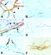
Early trophozoites in in vitro culture before exposure (A and D) and then 12 (B and E) and 24 hours (C and F) after exposure to IC99 concentrations of artemether (top row: A–C) and quinine (bottom row: D–F).
Figure 3.
Figure 3.
Brain vessels from four different cases of fatal falciparum malaria showing accumulations of different parasite stages. A, late trophozoites, B, schizonts with abundant pigment; C, mid-stage trophozoites; D; ring forms containing no intraerythrocytic pigment.
Figure 4.
Figure 4.
Brain vessels from four different cases of fatal falciparum malaria showing sequestration of intact schizonts (A and B), and free merozoites after schizont rupture (C and D). The intraparasitic malaria pigment has stained black in A and B, and the free pigment in C is colored brown-black.
Figure 5.
Figure 5.
Ring stage parasites in cerebral microvessels. 51: Accumulation of red cells containing ring form parasites in a cerebral vessel. The peripheral parasitemia at death was 0.9%. 52: Composite of two photomicrographs showing accumulation of ring stage parasitized red cells in a cerebral vessel. The peripheral parasite count was 0.2%. Both patients received quinine treatment. 53: Composite of vessels from four different patients (A–D), all of whom received quinine, showing ring stage accumulation.
Figure 6.
Figure 6.
Four different areas from the same patient showing vessels with (A) late trophozoites and early schizonts (A) ring stages (B) uninfected red cells (C) no malaria parasites but residual intravascular pigment (D). The patient was treated with artemether and died 50 hours after admission. The peripheral parasite count at death was 400/μl.
Figure 7.
Figure 7.
Composite photomicrograph (magnification, ×1000) of a brain smear from a fatal case of falciparum malaria. Four vessels are seen (A–D). Nearly all the red cells in A and B are parasitized, whereas few parasites are seen in C and D. A is intensely parasitized containing alternating clusters of trophozoites and schizonts. Residual adherent pigment in the far right of the vessel indicates earlier schizont rupture. In B younger trophozoites are seen evenly spaced on the left and schizonts on the right. The patient received artemether but died 4 hours later after the parasitemia had doubled from 14.5% to 34.9%.
Figure 8.
Figure 8.
Immunohistochemical staining of cerebral vessels from brain smears. A: Chondroitin sulfate staining from control case: note the focal sparse CS staining on endothelial cells compared to stronger staining on peri-vascular macrophages (arrows). B: Chondroitin sulfate staining on a vessels from a cases of cerebral malaria. Increased endothelial staining is seen (arrows) compared to control staining. C: Strong constitutive endothelial class I HLA staining. D: Negative CD36 staining on vessels from a malaria case.
Figure 9.
Figure 9.
Immunohistochemical staining in malaria-infected brain smears. A: Endothelial activation in a malaria infected brain as judged by increased ICAM-1 expression. An ICAM-1 positive vessel with associated sequestration (arrow) is shown with neighboring ICAM-1 positive vessels and no sequestration (arrowheads). B: Control case showing patchy resting ICAM-1 expression on cerebral vessels. C: Increased VCAM-1 staining in a malaria infected cases, showing variation in sequestration between neighboring vessels both with (arrow) and without (arrowheads) sequestration. D: Very low levels of patchy endothelial VCAM-1 staining in a control case. E: Patchy class II HLA staining on some endothelial cells unrelated to the presence of PRBC in one segment or over one endothelial cell. F: A dendritic astroglial cell from a smear of a cerebral malaria case showing strong class II HLA staining.
Figure 10.
Figure 10.
Brain vessel showing no malaria parasites but residual malaria pigment. The patient died after several days of quinine treatment. The peripheral parasite count at death was 80/μl.

References

    1. White NJ, Ho M: The pathophysiology of malaria. Adv Parasitol 1992, 31:83-173 - PubMed
    1. Clark IA, Rockett KA, Cowden WB: Possible central role of nitric oxide in conditions clinically similar to cerebral malaria. Lancet 1992, 340:894-896 - PubMed
    1. Clark IA, Rockett KA: The cytokine theory of cerebral malaria. Parasitol Today 1994, 10:410-412 - PubMed
    1. Berendt AR, Turner GDH, Newbold CI: Cerebral malaria: the sequestration hypothesis. Parasitol Today 1994, 10:412-414 - PubMed
    1. Udeinnya IJ, Schmidt JA, Aikawa M, Miller LM, Green I: Falciparum malaria infected erythrocytes specifically bind to cultured human endothelial cells. Science 1981, 213:555-557 - PubMed

Publication types

MeSH terms