. 1999 Aug;155(2):411-9.
doi: 10.1016/S0002-9440(10)65137-1.

Three-dimensional reconstruction of pulmonary arteries in plexiform pulmonary hypertension using cell-specific markers. Evidence for a dynamic and heterogeneous process of pulmonary endothelial cell growth

Affiliations

Three-dimensional reconstruction of pulmonary arteries in plexiform pulmonary hypertension using cell-specific markers. Evidence for a dynamic and heterogeneous process of pulmonary endothelial cell growth

C D Cool et al. Am J Pathol. 1999 Aug.

Abstract

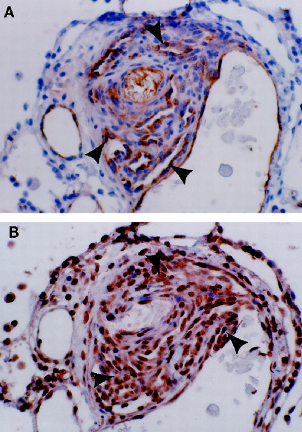
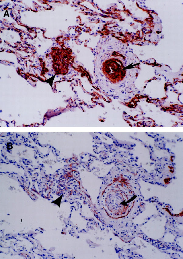
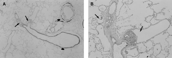
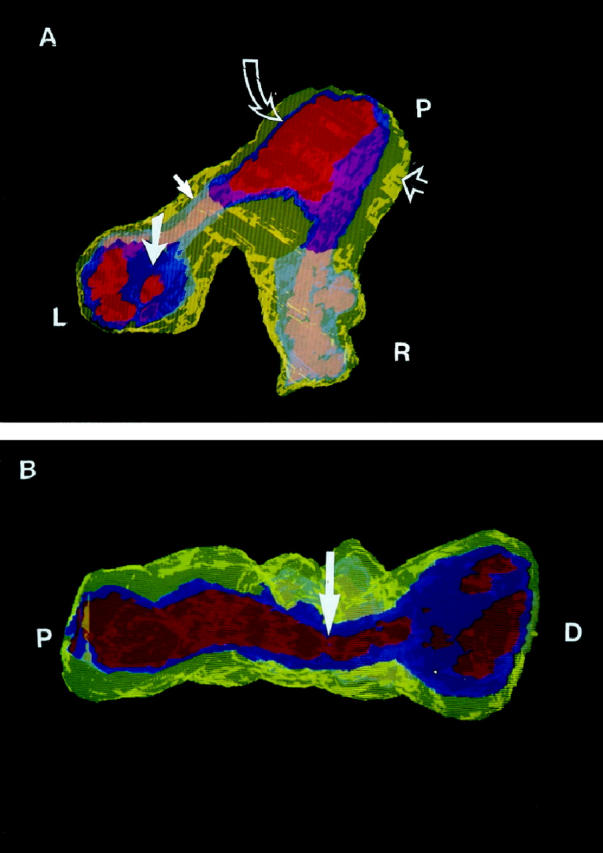
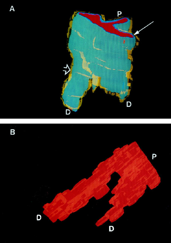
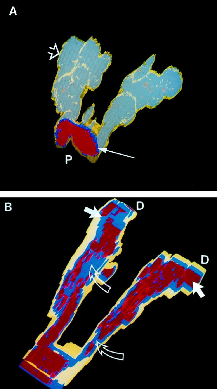
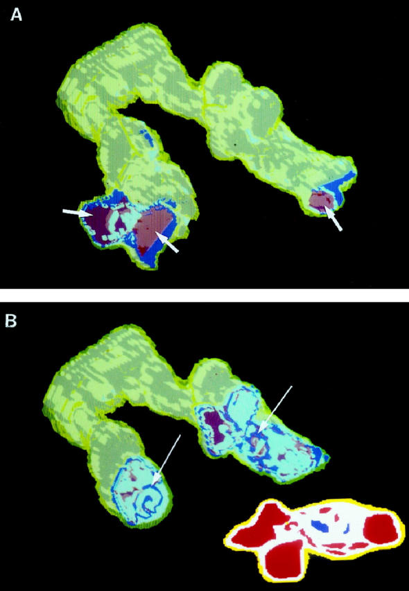
The plexiform lesions of severe pulmonary hypertension (PH) are complex vascular structures composed primarily of endothelial cells. In this study, we use immunohistochemical markers to identify the various cell layers of pulmonary vessels and to identify different endothelial cell phenotypes in pulmonary arteries affected by severe PH. Our computerized three-dimensional reconstructions of nine vessels in five patients with severe PH demonstrate that plexiform (n = 14) and concentric-obliterative (n = 6) lesions occur distal to branch points of small pulmonary arteries. And, whereas plexiform lesions occur as solitary lesions, concentric-obliterative lesions appear to be only associated with, and proximal to, plexiform structures. The endothelial cells of plexiform lesions express intensely and uniformly the vascular endothelial growth factor (VEGF) receptor KDR and segregate phenotypically into cyclin-kinase inhibitor p27/kip1-negative cells in the central core of the plexiform lesion and p27/kip1-positive cells in peripheral areas adjacent to incipient blood vessel formation. Using immunohistochemistry and three-dimensional reconstruction techniques, we show that plexiform lesions are dynamic vascular structures characterized by at least two endothelial cell phenotypes. Plexiform arteriopathy is not merely an end stage or postthrombotic change--it may represent one stage in an ongoing, angiogenic endothelial cell growth process.


Figures

Figure 1.
Figure 1.
Plexiform lesion from a patient with PPH. A: The multichanneled, cellular lesions stain positive for the FVIII-r.ag endothelial cell marker (arrowheads). B: Same lesion as A, immunostained for the VEGF receptor KDR. All of the endothelial cells stain positive for this endothelial cell-specific marker (arrowheads).
Figure 2.
Figure 2.
A: An illustration of the multiple concentric onion-skin pattern of the FVIII-r.ag-positive cells in a concentric-obliterative lesion (arrow). On the left is a conglomerate of positively stained endothelial cells, consistent with a partial slice through a plexiform lesion (arrowhead). B: For contrast, an adjacent section is stained for MSA. Note that the positive endothelial cells of the concentric-obliterative lesion do not stain with MSA (curved arrow). The surrounding smooth muscle coat is positive for MSA and negative for FVIII-r.ag. Also note that the plexiform lesion on the left is now more apparent in this deeper cut (arrowhead).
Figure 3.
Figure 3.
Longitudinal views of plexiform lesions in PPH. (A) Low-power magnification of a pulmonary artery stained for MSA. Distal to the bifurcation of the muscular artery, the two branching vessels are occluded by plexiform lesions (arrows). The thin smooth muscle layers of the bronchiolar and pulmonary arteries are highlighted by the MSA stain (short arrows). (B) Higher-power magnification of the same lesion stained with FVIII-r.ag. In this example, the endothelial cells lining the multiple lumina of the plexiform lesions stain positive for FVIII-r.ag. Dilatation lesions can be seen within and adjacent to the plexiform lesions (arrows).
Figure 4.
Figure 4.
(A) A three-dimensional cutaway view of plexiform and concentric-obliterative lesions in a bifurcating vessel from case 1 (PPH). Yellow, smooth muscle cells; ultramarine or gray-blue, endothelial cells; red or flesh-toned, lumen. (The apparent color changes are a result of the transparent overlying yellow smooth muscle and blue endothelial cell layers.) The proximal end (P) shows slight medial hypertrophy (yellow, open arrow), a normal endothelial cell monolayer (blue, curved open arrow), and an unobstructed lumen (red). At the left branch (L), the patent vessel turns into a concentric proliferation of endothelial cells (gray-blue, closed arrow), opening further distally into the multichanneled plexiform lesion (ultramarine, closed large arrow) . At the right branch (R), the patent vessel turns into a plexiform lesion (gray-blue). The blue endothelial cells of the plexiform lesions severely disrupt the lumina, best seen in (B). (B) A longitudinal view of the same vessel shows the proximal unobstructed lumen (P), a constriction distal to the branch point (arrow), and the proliferating and obstructing endothelial cells (blue) at the distal end (D) of one of the branches. This view highlights the transition of the patent pulmonary artery to concentric and plexiform changes.
Figure 5.
Figure 5.
(A) Three-dimensional view of a normal small pulmonary artery at a branch point (case 6). Note the thin yellow smooth muscle cell coat (open arrow) and the gray-blue endothelial cell monolayer (thin arrow). P, Proximal end; D, distal end. (B) Luminal cast of a normal vessel. Rotated view of A. Note the large, unobstructed channels, even distal to the branch point. P, Proximal end; D, distal end.
Figure 6.
Figure 6.
Lesion reconstructed from PPH case 2. (A) The gray-blue represents the endothelial cells stained with antibody directed against KDR. The yellow smooth muscle layer (open arrow) is thin, in marked contrast to PPH case 1 (Figure 4) ▶ . The distorted endothelial cell layers begin distal to the bifurcation of the vessel and can be seen through the translucent yellow smooth muscle coat. The proximal lumen (P) is lined by an endothelial cell monolayer (thin arrow). (B) Rotated and cutaway view of the same lesion. Note the severely disrupted red lumina distal to the bifurcation and the distal (D) dilatation lesions (arrows). Blue endothelial cells (open arrows) obstruct the lumina. Yellow, Smooth muscle; blue, KDR positive cells; red, lumen.
Figure 7.
Figure 7.
KDR and p27/kip1 staining pattern of the two plexiform lesions that are part of the complex vascular lesion depicted in Figure 6 ▶ . In these views, the distal portions of the arteries have been cut away. Yellow, Smooth muscle cells; red, lumen; white, KDR-positive/p27kip1-positive endothelial cells; blue, KDR-positive/p27/kip1-negative endothelial cells. (A) A view of the dilatation lesions (arrows) at the distal ends of the two pulmonary artery branches. (B) A more proximal cutaway view showing small foci of proliferating (p27/kip1-negative) endothelial cells (arrows). The inset shows a cross-sectional view of a two-dimensional map image used in the computerized reconstruction of case 2.

Comment in

References

    1. Wagenvoort CA, Wagenvoort N: Primary pulmonary hypertension. A histopathologic study of the lung vessels in 156 clinically diagnosed cases. Circulation 1970, 42:1163-1184
    1. Edwards, WD: Pathology of pulmonary hypertension. Cardiovasc Clin 1987, 18:321–354 - PubMed
    1. Saunders JB, Constable TJ, Heath D, Smith P, Paton A: Pulmonary hypertension complicating portal vein thrombosis. Thorax 1978, 34:281-283 - PMC - PubMed
    1. Robalino BD, Moodie DS: Association between primary pulmonary hypertension and portal hypertension: analysis of its pathophysiology and clinical, laboratory and hemodynamic manifestations. J Am Coll Cardiol 1991, 17:492-498 - PubMed
    1. Thomas SH, Butt AY, Corris PA, Egan JJ, Higenbottam TW, Madden BP, Waller PC: Appetite suppressants and primary pulmonary hypertension in the United Kingdom. Br Heart J 1995, 74:660-663 - PMC - PubMed

Publication types

MeSH terms