. 1999 Aug;155(2):461-72.
doi: 10.1016/S0002-9440(10)65142-5.

Distinct selectin ligands on colon carcinoma mucins can mediate pathological interactions among platelets, leukocytes, and endothelium

Affiliations

Distinct selectin ligands on colon carcinoma mucins can mediate pathological interactions among platelets, leukocytes, and endothelium

Y J Kim et al. Am J Pathol. 1999 Aug.

Abstract

Selectins are adhesion molecules that mediate calcium-dependent cell-cell interactions among leukocytes, platelets, and endothelial cells. The naturally occurring vascular ligands for the selectins are mostly mucin-type glycoproteins. Increased expression and altered glycosylation of mucins are known to be prominent features of carcinoma progression. We have previously shown that all three selectins bind to colon carcinoma cell lines in a calcium-dependent fashion and that carcinoma growth and metastasis formation are attenuated in P-selectin-deficient mice. Here we show that the three recombinant soluble selectins recognize ligands within primary colon carcinoma tissue samples. Affinity chromatography showed that the ligands for all three selectins are O-sialoglycoprotease-sensitive mucins that are recognized in a calcium- and sialic acid-dependent manner. Furthermore, there are separate binding sites on the mucins for each selectin, allowing cross-binding of a single mucin molecule by more than one selectin. We also show that the selectin ligands on purified carcinoma mucins can mediate at least four different pathological interactions among platelets, leukocytes, and endothelial cells. These findings could explain some of the adhesive events of blood-borne tumor cells reported to occur with leukocytes, platelets, and endothelial cells, which are believed to play a part in modulating some early events in tumor metastases.


Figures
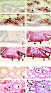
Figure 1.
Figure 1.
Heterogeneous expression of selectin ligands in human colon carcinoma samples. Frozen (upper panel) or paraffin (middle and lower panels) sections of primary human colon carcinomas (upper and middle panels) or normal colon (lower panel) were stained with the recombinant soluble FLAG-epitope tagged selectin probes in the presence of calcium as described in Materials and Methods. Each panel shows a similar region of one of the samples studied: L, L-selectin-FLAG-Rg; E, E-selectin-FLAG-Rg; P, P-selectin-FLAG-Rg. In each panel, the control (C) shows an example of loss of binding on incubation of E-selectin-Rg in EDTA-containing buffer.
Figure 2.
Figure 2.
Interactions of purified carcinoma mucins with recombinant selectins. A: Lack of competition between carcinoma mucins for binding to each of the selectins. Fixed amounts of each biotinylated selectin were mixed with serial dilutions of the three unlabeled selectins before incubation on the ELISA wells coated with LS180 mucins. Saturation curves were generated for each biotinylated selectin (data not shown); the concentrations used (2 μg/ml of biotinylated E- and P-selectins and 1 μg/ml of biotinylated L-selectin) were within the linear range of detection response. Each panel displays the binding of the biotinylated selectins in the presence of increasing amounts of unlabeled selectins. The percentage inhibition values were calculated after EDTA control subtraction in comparison to wells that contained only the relevant biotinylated selectin. B: Mucins captured by each of the three selectins contain additional binding sites. Microwell plates were coated with each of the three recombinant selectin-Igs before blocking and incubation with carcinoma mucins. After washing FLAG-epitope-tagged selectin-Ig molecules precomplexed with anti-FLAG antibody-alkaline phosphatase were added, incubated, washed again, and developed as described in Materials and Methods. The data shown are representative of four experiments that yielded similar results. Background readings obtained in presence of EDTA have been subtracted.
Figure 3.
Figure 3.
Carcinoma mucins potentiate platelet aggregation. Freshly prepared human platelets were studied for aggregation in a platelet aggregometer after various dosages of thrombin and/or mucin. The results shown are representative of several experiments. A: ∼20 × 10 human platelets were initially incubated in the presence of 0.11 μg/uL of carcinoma mucins without any effect. When 0.5 U of human thrombin was added thereafter, maximal aggregation was noted. Without mucins (buffer only), the same amount of thrombin was suboptimal, giving only partial aggregation. B: Mucins and thrombin were added together at the same concentrations as in A. Mucin potentiation of aggregation was inhibited by a blocking anti-P-selectin antibody (C138A), but not with a nonblocking P-selectin antibody (S12). C: The reverse order of addition gives similar results; mucin added after suboptimal dose of thrombin induced further aggregation.
Figure 4.
Figure 4.
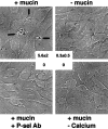
Carcinoma mucins cause interactions of activated platelets with activated endothelium. Human platelets (5 × 10⁶) were added to HUVEC cells in a 6-well plate with or without LS180 carcinoma mucins (40 μg/ml) or thrombin (0.1 U/well) as described in Material and Methods. No aggregation or attachment of platelets occurred in the absence of a thrombin activation, regardless of whether mucin was added (data not shown) In all panels shown, both HUVEC cells and platelets were activated with thrombin. The upper panels show examples (arrows) of aggregates of platelets attached to the HUVEC cells in the presence of mucins and a marked decrease in the absence of mucin. Aggregates were essentially undetectable even with mucin added in the presence of a P-selectin-blocking antibody or in the absence of calcium ions. The numbers indicate the number of aggregates seen per high-power field (mean ± SD of 10 fields).
Figure 5.
Figure 5.
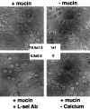
Carcinoma mucins agglutinate peripheral blood leukocytes in a calcium- and L-selectin-dependent manner. Human PBMCs were washed and incubated with or without carcinoma mucins (40 μg/ml) as described in Materials and Methods. Examples are shown of the agglutination observed in the presence of mucins, which was markedly reduced by calcium chelation with 1 mmol/L EDTA, or by adding a blocking anti-L-selectin antibody. The numbers indicate the number of leukocyte aggregates of >5 cells seen per high-power field (mean ± SD of 10 fields).
Figure 6.
Figure 6.
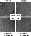
Carcinoma mucins mediate interactions of human PBMCs with activated endothelium via L- and E-selectin. 1 × 10 washed PBMCs were added to HUVECs grown in a 6-well plate that had been previously incubated for 4 hours in complete medium containing 500 μg/ml of TNF-α and washed once in the interaction buffer (see Material and Methods). The incubations were carried out with or without carcinoma mucins (40 μg/ml) as indicated with gentle shaking at room temperature for 10 minutes, and the wells washed three times in the same buffer. The binding of the PBMCs to the HUVECs was enhanced by the presence of mucin, markedly reduced by an E-selectin-blocking antibody, and partially reduced by an L-selectin-blocking antibody (to about the level seen without mucin). The numbers indicate the number of attached leukocytes seen per high-power field (mean ± SD of 10 fields). PBMC binding was almost completely absent if the HUVECs’ pre-incubation did not include TNF-α or, in the absence of calcium ions, regardless of whether carcinoma mucins were present (data not shown).
Figure 7.
Figure 7.
Potential interactions between vascular selectins and the multiple binding sites on colon carcinoma mucins. The composite schematic shows potential interactions of cell surface or secreted carcinoma mucins with selectins expressed on leukocytes, platelets, or endothelium; it was assumed that activation of endothelium and/or platelets could occur in the setting of a systemic malignancy. Although all of these interactions may not occur at the same time, multiple interactions could potentially occur in various combinations. Several of these have been demonstrated in this study using purified carcinoma mucins. Interactions involving the natural endogenous cell surface E- and P-selectin ligands, present on many leukocytes, are not shown.

References

    1. Rosen SD, Bertozzi CR: The selectins and their ligands. Curr Opin Cell Biol 1994, 6:663-673 - PubMed
    1. Lasky LA: Selectin-carbohydrate interactions and the initiation of the inflammatory response. Annu Rev Biochem 1995, 64:113-139 - PubMed
    1. Ley K, Tedder TF: Leukocyte interactions with vascular endothelium: New insights into selectin-mediated attachment and rolling. J Immunol 1995, 155:525-528 - PubMed
    1. Nelson RM, Venot A, Bevilacqua MP, Linhardt RJ, Stamenkovic I: Carbohydrate-protein interactions in vascular biology. Annu Rev Cell Biol 1995, 11:601-631 - PubMed
    1. Tedder TF, Steeber DA, Chen A, Engel P: The selectins: vascular adhesion molecules. FASEB J 1995, 9:866-873 - PubMed

Publication types

MeSH terms

LinkOut - more resources