. 1999 Apr;83(4):414-9.
doi: 10.1136/bjo.83.4.414.

Allo-limbal transplantation in patients with limbal stem cell deficiency

Affiliations

Allo-limbal transplantation in patients with limbal stem cell deficiency

H S Dua et al. Br J Ophthalmol. 1999 Apr.

Abstract

Aim: To report the outcome of a series of patients with stem cell deficiency who underwent allo-limbal transplantation and to describe a technique for this procedure.

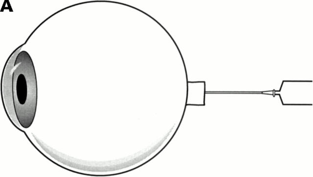
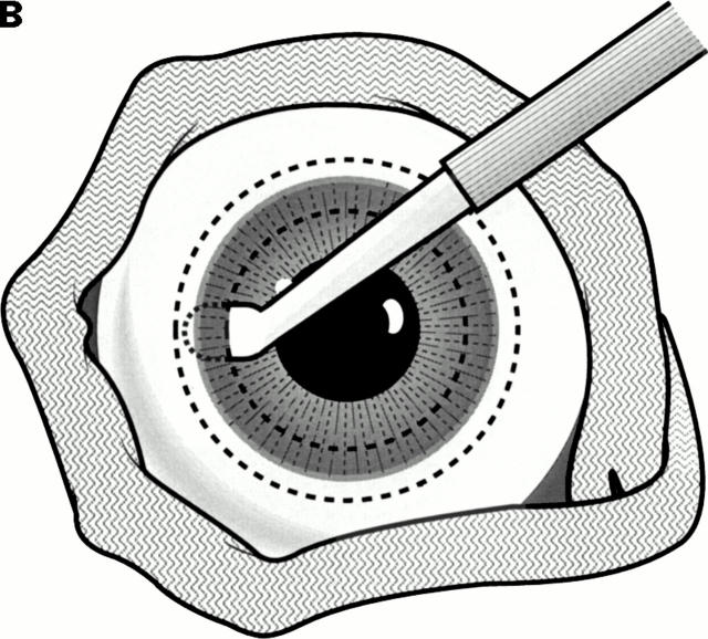
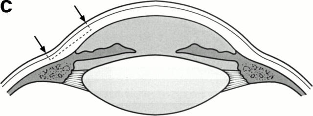
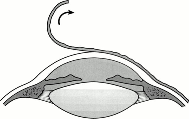
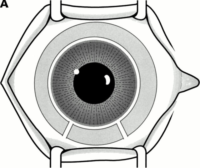
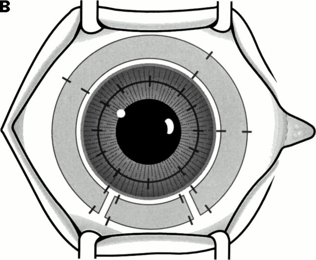
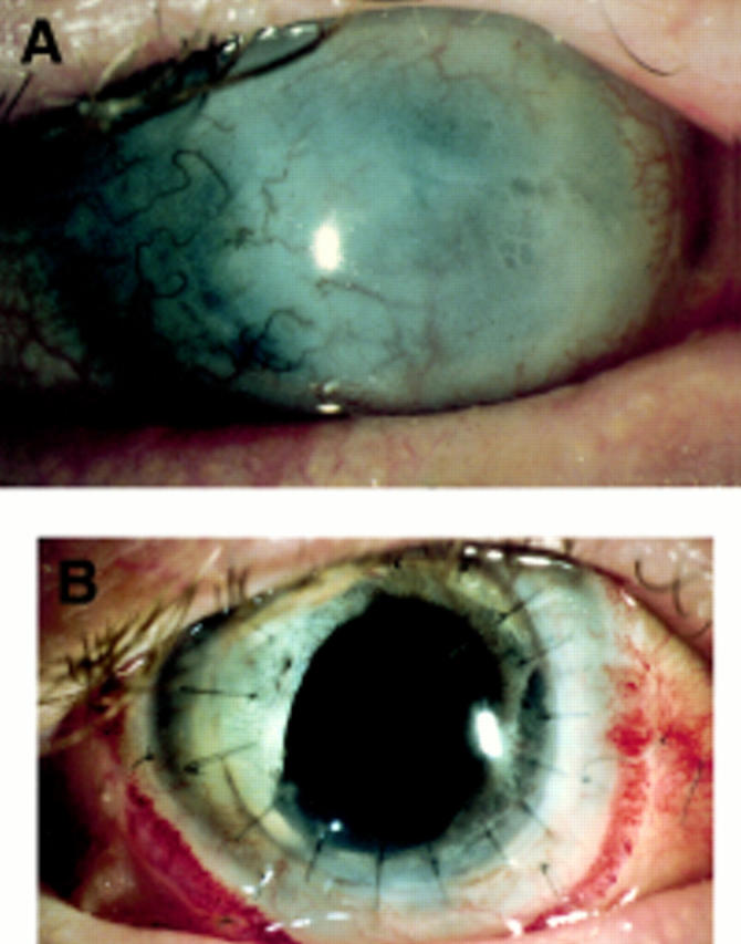
Methods: Six consecutive patients underwent allo-limbal stem cell transplantation. The primary diagnosis included alkali burn (n = 2), trachoma (n = 1), chronic rosacea blepharitis and kerato-conjunctivitis (n = 1), aniridia (n = 1), and Stevens-Johnson syndrome (n = 1). The limbal rim consisted of peripheral cornea and perilimbal sclera. FK-506 was used postoperatively for immunosuppression.

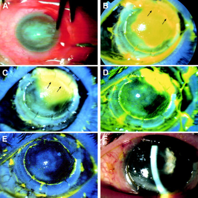
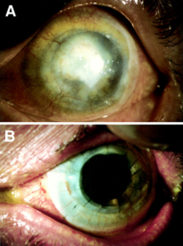
Results: The length of follow up ranged from 3 to 24 months (mean follow up 11.8 (SD 9.3) months). The outcome was considered satisfactory in five of six cases. The corneal surface was completely epithelialised within 2 weeks, and there was a substantial improvement in vision and symptoms. One patient had recurrent epithelial defects related to eyelid abnormalities. No side effects associated with systemic immunosuppression were noted.

Conclusion: Allo-limbal transplantation, with systemic immunosuppression with FK-506 is useful in reconstruction of the ocular surface with improvement in vision in patients with severe stem cell deficiency.


Figures

Figure 1
Figure 1
Surgical technique. Harvesting donor limbus. (A) Inflating eyeball with sterile air injected through the optic nerve. This route allows air to remain trapped in the vitreous cavity and keeps the eye firm during tissue dissection. (B) Lamellar dissection of a ring of peripheral cornea, limbus, and adjacent sclera with an angled bevel up blade. The donor eyeball is wrapped with a strip of gauze to facilitate handling. (C) The diagram illustrates the location of tissue dissected.
Figure 1
Figure 1
Surgical technique. Harvesting donor limbus. (A) Inflating eyeball with sterile air injected through the optic nerve. This route allows air to remain trapped in the vitreous cavity and keeps the eye firm during tissue dissection. (B) Lamellar dissection of a ring of peripheral cornea, limbus, and adjacent sclera with an angled bevel up blade. The donor eyeball is wrapped with a strip of gauze to facilitate handling. (C) The diagram illustrates the location of tissue dissected.
Figure 2
Figure 2
Surgical technique. Preparing the host bed. After a 360° peritomy, a plane of dissection superficial to corneal stroma is created and the entire fibrovascular membrane is lifted off the surface.
Figure 3
Figure 3
Surgical technique. Transplanting the donor limbal ring. (A) The harvested ring of tissue carrying the "stem cells" is placed around the recipient limbus. A small arc of similar tissue, to fill the gap between the cut ends of the limbal ring, is cut to size and appropriately placed. (B) The limbal explants are sutured along both inner and outer circumferences using 10-0 nylon sutures. The sutures are placed directly opposite to each other. In this illustration a central penetrating keratoplasty is also shown.
Figure 3
Figure 3
Surgical technique. Transplanting the donor limbal ring. (A) The harvested ring of tissue carrying the "stem cells" is placed around the recipient limbus. A small arc of similar tissue, to fill the gap between the cut ends of the limbal ring, is cut to size and appropriately placed. (B) The limbal explants are sutured along both inner and outer circumferences using 10-0 nylon sutures. The sutures are placed directly opposite to each other. In this illustration a central penetrating keratoplasty is also shown.
Figure 4
Figure 4
Results of limbal transplant in case 2 (primary diagnosis: aniridia). (A) The external appearance of the right eye showing an extensive fibrovascular membrane covering the cornea. The picture was taken during surgery while a peritomy was being done. (B) Slit lamp photograph (fluorescein stained) of the same eye 3 days after surgery. Corneal epithelial cells have started to migrate from the transplanted limbus and are migrating across the host cornea. The quadrant indicated with arrows, where no epithelial cell migration is seen, corresponds to the site where a spacer, fashioned from corneal collagen (without epithelial cover), was placed. (C) Slit lamp photograph (fluorescein stained) of the same eye, 5 days after surgery. Three quarters of the host surface is re-epithelialised. The quadrant corresponding to the spacer (arrows) has remained bare. (D) Slit lamp photograph (fluorescein stained) of the same eye, 7 days after surgery. The remaining quadrant is almost covered by cells migrating from the other quadrants. (E) Slit lamp photograph (fluorescein stained) of the same eye taken on day 8 post surgery. Re-epithelialisation of the host corneal surface from donor limbus is complete. (F) Slit lamp photograph of the same eye 6 weeks after surgery. The cornea is clear and the surface is smooth as illustrated by the broad slit beam. Remnants of the lens capsule (following needling for congenital cataract) with a central opening are now clearly visible.
Figure 5
Figure 5
Results of combined limbal and corneal transplant with lens extraction and implant (quadruple procedure) in case 3. (A) Preoperative picture of the left eye of a patient with clinical diagnosis of trachoma of 30 years' duration. The cornea is scarred and vascularised with calcium deposition. The outline of a previously failed corneal graft is visible. Preoperative vision was hand movements. (B) The same eye 8 weeks after surgery. The patient has a follow up of 9 months and maintains a vision of 6/12 unaided and 6/9 with a pinhole.
Figure 6
Figure 6
Results of combined limbal and corneal transplant with lens extraction and implant (quadruple procedure) in case 6. (A) Preoperative picture of the right eye of a patient with clinical diagnosis of Stevens-Johnson syndrome. The cornea is scarred and vascularised. Preoperative vision was light perception. (B) The same eye 4 weeks after surgery.

Comment in

References

    1. Am J Ophthalmol. 1974 Apr;77(4):538-42 - PubMed
    1. Ophthalmology. 1995 Jan;102(1):101-7 - PubMed
    1. Am J Ophthalmol. 1984 Jan;97(1):1-6 - PubMed
    1. J Cell Biol. 1986 Jul;103(1):49-62 - PubMed
    1. Cornea. 1984-1985;3(4):231-9 - PubMed