. 1999 Apr;83(4):420-4.
doi: 10.1136/bjo.83.4.420.

Assessment of macular function by multifocal electroretinogram before and after macular hole surgery

Affiliations

Assessment of macular function by multifocal electroretinogram before and after macular hole surgery

Y J Si et al. Br J Ophthalmol. 1999 Apr.

Abstract

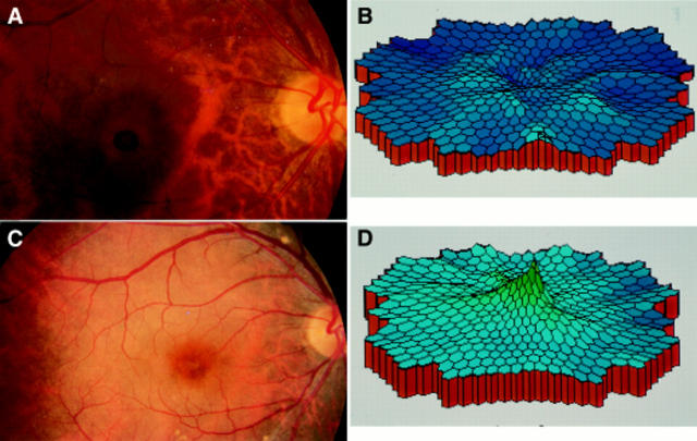
Aim: To evaluate macular function before and after successful surgical closure of idiopathic macular holes using multifocal electroretinogram (ERG).

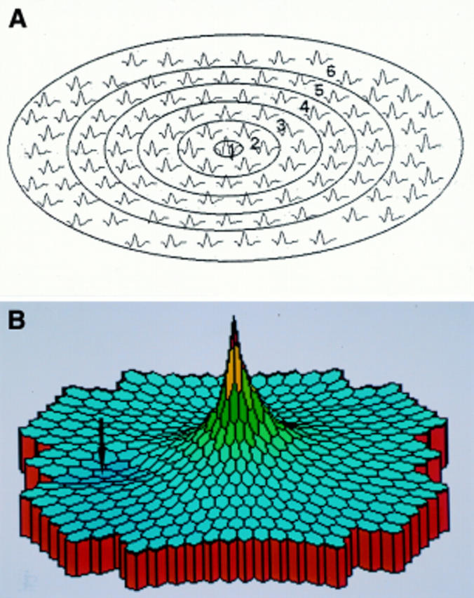
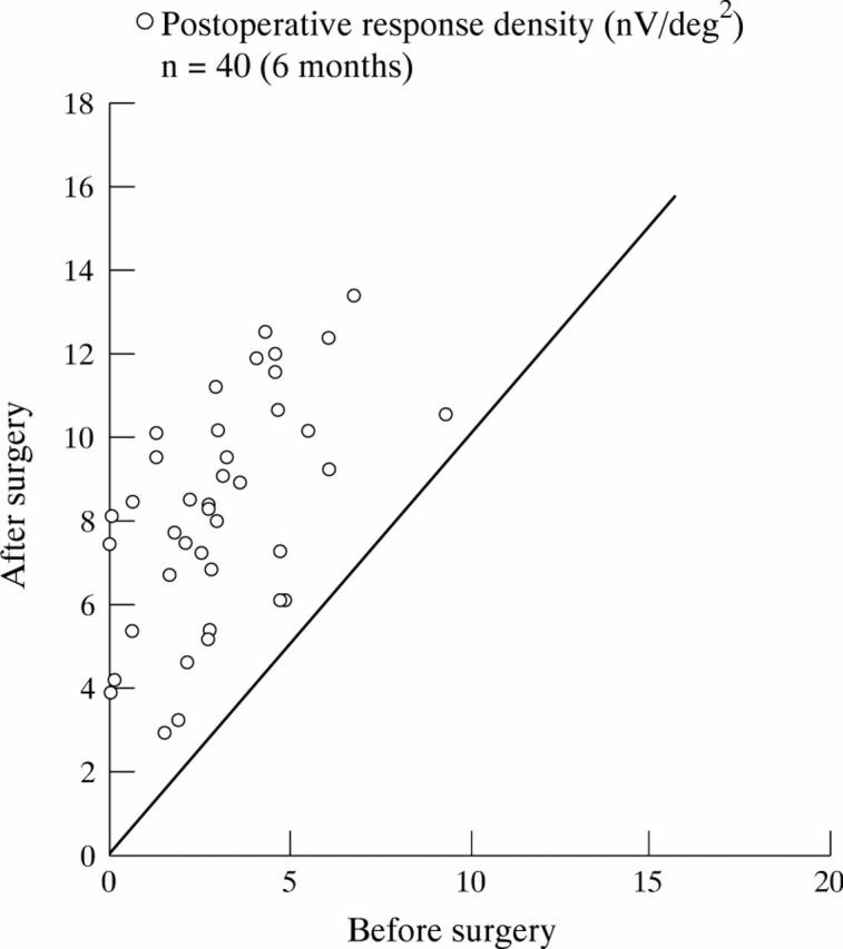
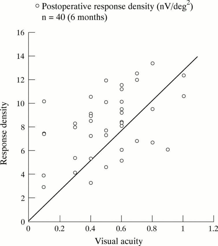
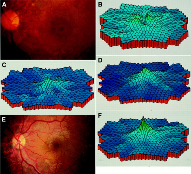
Methods: 40 patients (40 eyes) with idiopathic macular holes were examined using multifocal ERG both before and after vitreous surgery. The postoperative period was from 1 to 12 months.

Results: Preoperatively, the electrical retinal response densities in the foveal and the perifoveal area were apparently decreased. After a mean postoperative period of 3-6 months, the foveal and perifoveal area electrical retinal response densities improved to two to four times the preoperative level and the improvement continued to 1 year after surgery.

Conclusion: In macular holes, the decrease in retinal electrophysiological response was not limited to the fovea but involved an area of the perifovea of 1.6 disc diameters. The electrical retinal response density of these areas gradually improved after macular hole closure.


Figures

Figure 1
Figure 1
Schema of the stimulus matrix of multifocal ERG (A), and three dimensional scalar plots of ERG responses obtained from the left eye of a normal subject (B). There is a sharp peak at the foveal area and slight depression at the optic disc (arrow).
Figure 2
Figure 2
Relation between preoperative and postoperative retinal response density 6 months after surgery. Retinal response density increased in all eyes (p<0.001).
Figure 3
Figure 3
Association between visual acuity and the retinal response density 6 months after surgery. A positive correlation between response density and visual acuity is seen (p<0.01). Retinal response density varied in eyes with the same visual acuity.
Figure 4
Figure 4
Multifocal ERG of a 75 year old man with a stage 3 macular hole (case 1, left eye) before and after surgery. Preoperative fundus photograph (A) and multifocal ERG topography (B). Foveal and perifoveal area (areas 1 and 2) showed crater like depression. Multifocal ERG recorded at 1 month (C) and 3 months (D) postoperatively. The peak recovered in the macular area. Fundus photograph (E) and multifocal ERG topography (F) 6 months postoperatively.
Figure 5
Figure 5
Fundus photograph and multifocal ERG of a 62 year old man with a stage 3 macular hole (case 2, right eye) before and after surgery. (A) Fundus photograph preoperatively. Macular hole is larger than that of the patient in case 1. (B) Multifocal ERG topography showed more widespread crater-like depression in macular area than that in case 1. Fundus photograph (C) and multifocal ERG topography (D) 6 months postoperatively. Macular hole had closed. The peak of the retinal response density was restored in macular area in the topography of multifocal ERG.

References

    1. Invest Ophthalmol Vis Sci. 1986 Jul;27(7):1123-30 - PubMed
    1. Doc Ophthalmol. 1996-1997;92(4):291-300 - PubMed
    1. Ophthalmology. 1987 Sep;94(9):1134-9 - PubMed
    1. Arch Ophthalmol. 1988 May;106(5):629-39 - PubMed
    1. Ophthalmology. 1988 Jul;95(7):917-24 - PubMed