Case Reports
. 1999 Apr;83(4):452-7.
doi: 10.1136/bjo.83.4.452.

Lens epithelial changes and mutated gene expression in patients with myotonic dystrophy

Affiliations
Case Reports

Lens epithelial changes and mutated gene expression in patients with myotonic dystrophy

T Abe et al. Br J Ophthalmol. 1999 Apr.

Abstract

Aims: Examination of the expression of the mutated allele of myotonic dystrophy protein kinase gene and lens epithelial cell changes in patients with myotonic dystrophy.

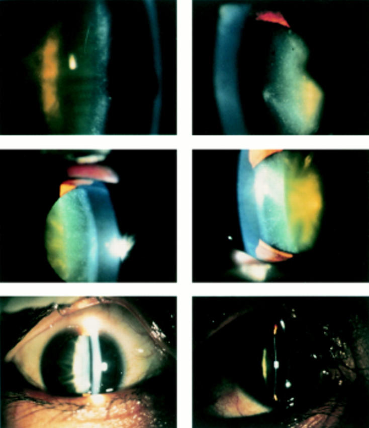
Methods: Six eyes from three patients with myotonic dystrophy underwent cataract surgery. The lens epithelium was photographed to examine the morphological changes. mRNAs were extracted to determine myotonic dystrophy protein kinase gene expression in the lens epithelium and peripheral blood. Age matched lens epithelial cells from senile cataracts were used as controls.

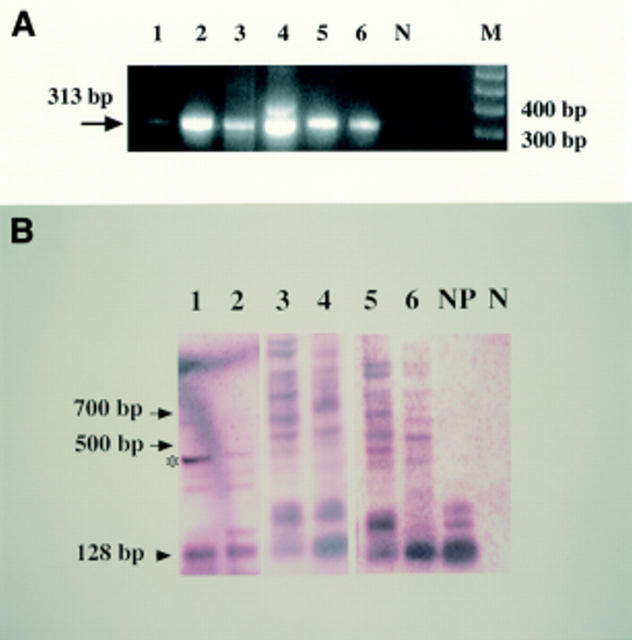
Results: All eyes showed iridescent or posterior subcapsular lens opacity. The expression of the myotonic dystrophy protein kinase gene with trinucleotide repeat expansion was evaluated by reverse transcriptase polymerase chain reaction, Southern blotting, and sequence analysis. Lens epithelial cell densities were extremely reduced in the patients compared with the control group.

Conclusion: To the authors' knowledge, this is the first report to describe the relation between lens epithelial cell changes and mutated gene expression in patients with myotonic dystrophy. The gene may be mitotically unstable in the lens epithelial cells; it may influence cell density and lens epithelial function, and it may lead to the development of typical subcapsular lens opacity.


Figures

Figure 1
Figure 1
Pedigrees of three families showing generations (roman numerals) of affected (closed symbols) and unaffected (open symbols) members. Squares indicate male members; circles, female; and slash, deceased.
Figure 2
Figure 2
Photographs of cataracts were taken in affected patients. Patient H-II-2 (top, right and left are right eye and left eye, respectively) is a 59 year old man; patient T-II-4 (middle; right and left eyes) is a 58 year old woman; and patient O-III-1 (bottom; right and left eyes) is a 33 year old man. All showed either cortical iridescent opacity or posterior cortical opacity. Patients are described in the text.
Figure 3
Figure 3
Examination of lens epithelial cell density reveals that all MD patients with cataract show extremely decreased cell densities, compared with the age matched normal subject. Lens epithelium of patient T-II-4 (top; right and left are right eye and left eye, respectively), a 58 year old woman; patient H-II-2 (second from top; right and left eyes), a 59 year old man; patient O-III-1 (second from bottom; right and left eyes), a 33 year old man; control subject with senile cataract (bottom right), a 78 year old man and also other control subject with senile cataract (bottom left), a 58 year old man. Actual cell density is shown in Table 1.
Figure 4
Figure 4
Results of reverse transcriptase polymerase chain reaction (RT-PCR) and Southern blotting analysis from affected patients in the families. (A) Results of β actin from lens epithelial cells and peripheral blood from patients. Patient H-II-2 (Nos 1 and 2 indicate lens epithelial cells and peripheral blood, respectively) is a 59 year old man; patient T-II-4 (Nos 3 and 4, same as above) is a 58 year old woman; and patient O-III-1 (Nos 5 and 6, same as above) is a 33 year old man. The negative control is indicated by N. M is a marker of the 100 base pair ladder. (B) Results of myotonic dystrophy protein kinase gene. Normal band is indicated by arrowhead; extended bands, arrows. The normal control (NP) show no mutated band; conversely, mutated bands occur in all affected members examined. The extended band, which was shown by an asterisk, was subcloned and sequenced.
Figure 5
Figure 5
Result of the sequence of the subcloned DNA, which was amplified from the cDNA of the lens epithelium in patient T-II-4 (shown by an asterisk in Fig 4B[f4}. Abnormal CAG repeat, the reverse sequence of CTG, was observed in the amplified DNA.

References

    1. Ophthalmology. 1984 Oct;91(10):1135-40 - PubMed
    1. J Med Genet. 1995 Aug;32(8):593-6 - PubMed
    1. Muscle Nerve. 1994 Sep;17(9):1037-43 - PubMed
    1. Graefes Arch Clin Exp Ophthalmol. 1996 Jul;234(7):419-24 - PubMed
    1. Cell. 1992 Feb 21;68(4):799-808 - PubMed

Publication types

Substances