

doi: 10.1091/mbc.10.8.2475.
Compartmentalization of the erythrocyte membrane by the membrane skeleton: intercompartmental hop diffusion of band 3
Affiliations
- PMID: 10436005
- PMCID: PMC25476
- DOI: 10.1091/mbc.10.8.2475
Item in Clipboard
Compartmentalization of the erythrocyte membrane by the membrane skeleton: intercompartmental hop diffusion of band 3
Mol Biol Cell.
1999 Aug.
Free PMC article
No abstract available
Figures
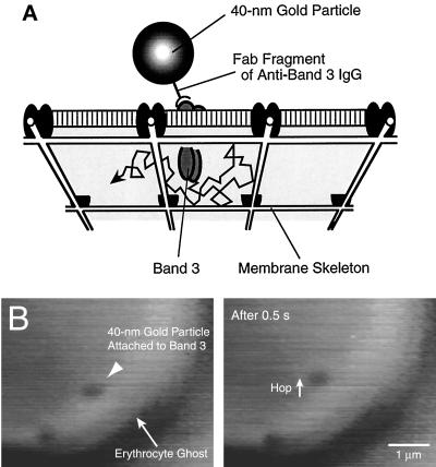
(A) The model to explain the confined diffusion of band 3 in the membrane (the membrane skeleton fence model). (B) Movement of gold particles attached to band 3 in the erythrocyte membrane, observed with a temporal resolution of 8 ms (4 times greater than the normal video rate).
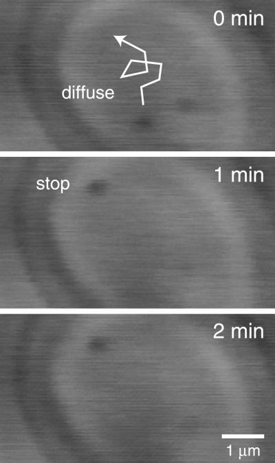
Long-term observations of the movement of band 3 in the erythrocyte membrane. Images recorded every 250 ms using time-lapse video microscopy.
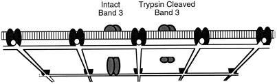
Movement of trypsin-cleaved band 3 was recorded with temporal resolutions of 8 and 0.89 ms.
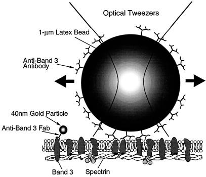
Deformation of the membrane skeleton using optical tweezers. A 1-μm latex bead, coated with anti-band 3 IgG, bound multiple band 3 molecules, of which ∼30% are attached to the membrane skeleton. By dragging the latex bead, it was possible to deform the membrane skeleton, which can be observed by single particle tracking using gold particles specifically attached to spectrin on the cytoplasmic surface of the erythrocyte ghost membrane.
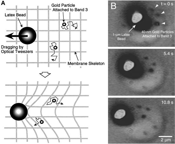
(A) Effect of lateral dragging of a 1-μm latex bead attached to the membrane skeleton on the movement of several band 3 molecules capable of undergoing hop diffusion. (B) The gold particles bound to band 3 undergoing hop diffusion followed the bead when it was dragged toward the left at a rate of 0.15 μm/s.
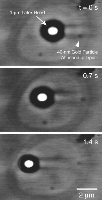
Effect of lateral dragging of a 1-μm latex bead attached to the membrane skeleton on the movement of a lipid that is located in the outer leaflet of the cell membrane.
Similar articles
-
Regulation mechanism of the lateral diffusion of band 3 in erythrocyte membranes by the membrane skeleton.J Cell Biol. 1998 Aug 24;142(4):989-1000. doi: 10.1083/jcb.142.4.989. J Cell Biol. 1998. PMID: 9722611 Free PMC article.
-
Anion exchanger 1 (band 3) is required to prevent erythrocyte membrane surface loss but not to form the membrane skeleton.Cell. 1996 Sep 20;86(6):917-27. doi: 10.1016/s0092-8674(00)80167-1. Cell. 1996. PMID: 8808627
-
Cytoskeleton-membrane connections in the human erythrocyte membrane: band 4.1 binds to tetrameric band 3 protein.Biochim Biophys Acta. 1997 Apr 26;1325(2):226-34. doi: 10.1016/s0005-2736(96)00261-1. Biochim Biophys Acta. 1997. PMID: 9168148
-
Red cell membrane cytoskeleton and the control of membrane properties.Biochem Soc Trans. 1991 Nov;19(4):1039-41. doi: 10.1042/bst0191039. Biochem Soc Trans. 1991. PMID: 1794461 Review. No abstract available.
-
[Band 3 protein, anion exchange protein of the erythrocyte membrane].Nihon Rinsho. 1992 Sep;50(9):2069-76. Nihon Rinsho. 1992. PMID: 1433993 Review. Japanese.
Cited by
-
Imaging of the diffusion of single band 3 molecules on normal and mutant erythrocytes.Blood. 2009 Jun 11;113(24):6237-45. doi: 10.1182/blood-2009-02-205450. Epub 2009 Apr 15. Blood. 2009. PMID: 19369229 Free PMC article.
-
Chemical biology approaches to membrane homeostasis and function.Chimia (Aarau). 2011;65(11):849-52. doi: 10.2533/chimia.2011.849. Chimia (Aarau). 2011. PMID: 22289370 Free PMC article. Review.
-
Nanoscopic compartmentalization of membrane protein motion at the axon initial segment.J Cell Biol. 2016 Oct 10;215(1):37-46. doi: 10.1083/jcb.201603108. Epub 2016 Oct 3. J Cell Biol. 2016. PMID: 27697928 Free PMC article.
-
Soft picture of lateral heterogeneity in biomembranes.J Membr Biol. 2003 Nov 15;196(2):135-46. doi: 10.1007/s00232-003-0633-z. J Membr Biol. 2003. PMID: 14724750
-
Single molecule imaging of green fluorescent proteins in living cells: E-cadherin forms oligomers on the free cell surface.Biophys J. 2001 Jun;80(6):2667-77. doi: 10.1016/S0006-3495(01)76236-4. Biophys J. 2001. PMID: 11371443 Free PMC article.
References
-
- Bennett V. Spectrin-based membrane skeleton: a multipotential adaptor between plasma membrane and cytoplasm. Physiol Rev. 1990;70:1029–1065. - PubMed
-
- Bennett V, Gilligan DM. The spectrin-based membrane skeleton and micron-scale organization of the plasma membrane. Annu Rev Cell Biol. 1993;9:27–66. - PubMed
-
- Edidin M, Kuo SC, Sheetz MP. Lateral movements of membrane glycoproteins restricted by dynamic cytoplasmic barriers. Science. 1991;254:1379–1382. - PubMed
-
- Golan DE. Red blood cell membrane protein and lipid diffusion. In: Agre P, Parker JC, editors. Red Blood Cell Membranes. New York: Marcel Dekker; 1989. pp. 367–400.
-
- Jacobson K, Sheets ED, Simson R. Revisiting the fluid mosaic model of membranes. Science. 1995;268:1441–1442. - PubMed
MeSH terms
Substances
LinkOut - more resources
Full Text Sources
