. 1999 Aug;10(8):2607-18.
doi: 10.1091/mbc.10.8.2607.

The spindle checkpoint of budding yeast depends on a tight complex between the Mad1 and Mad2 proteins

Affiliations
Free PMC article

The spindle checkpoint of budding yeast depends on a tight complex between the Mad1 and Mad2 proteins

R H Chen et al. Mol Biol Cell. 1999 Aug.
Free PMC article

Abstract

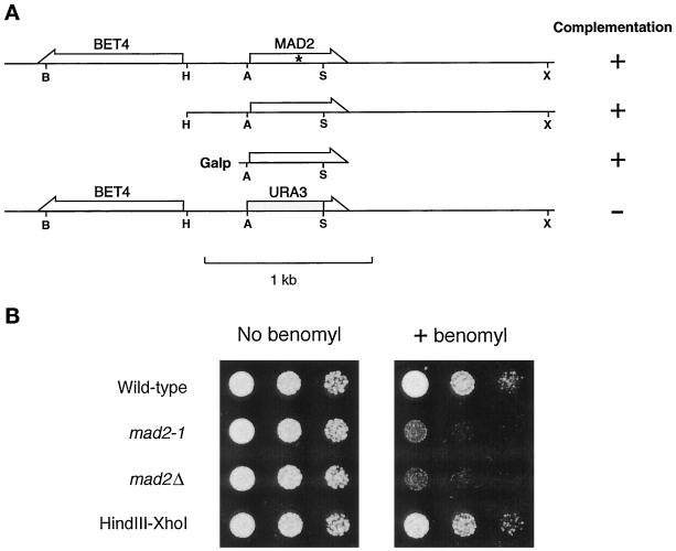
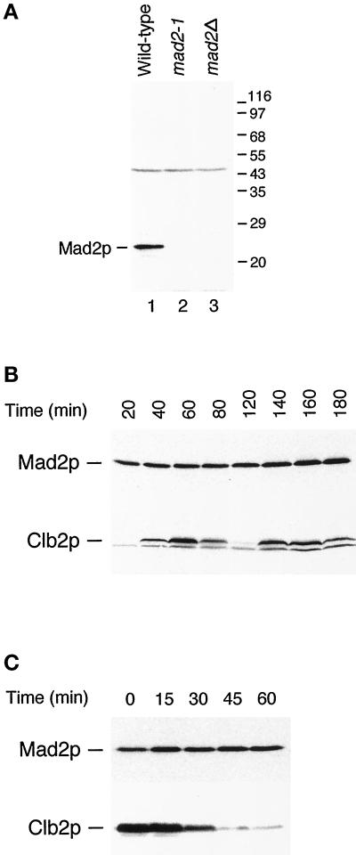
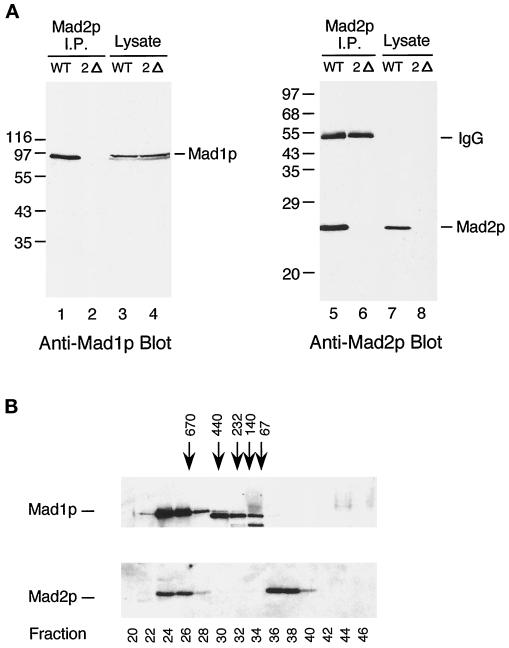
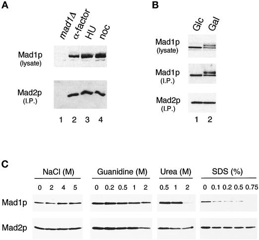
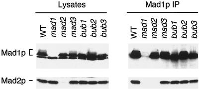
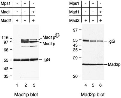
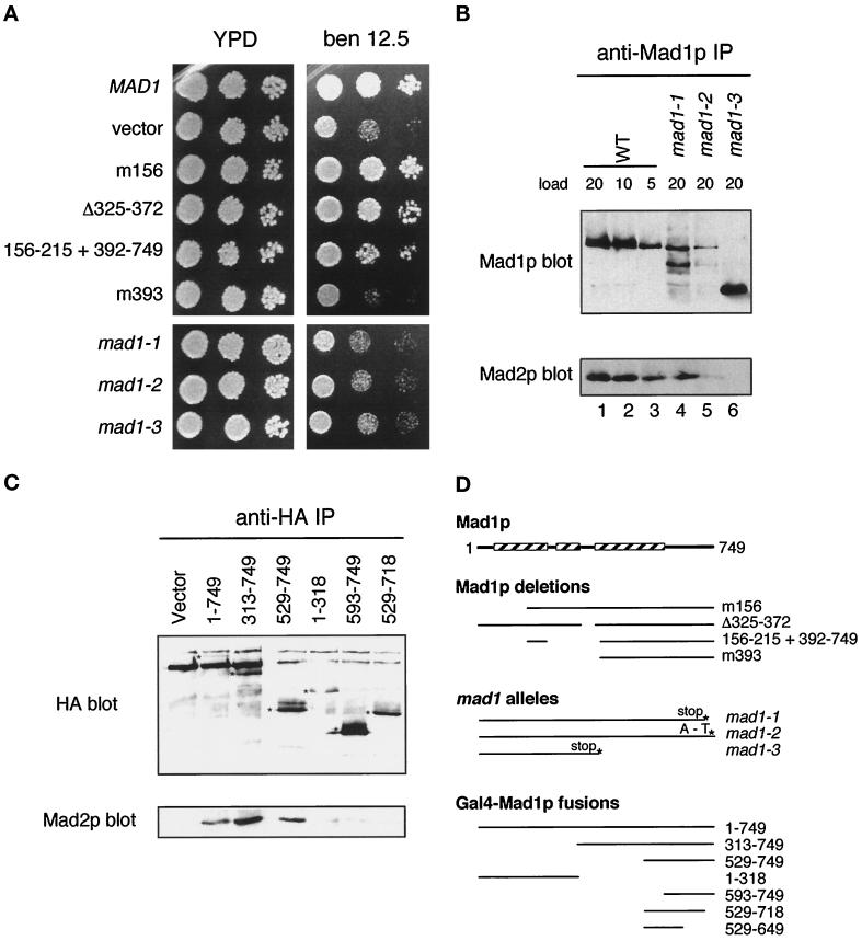
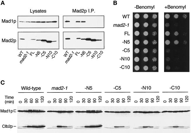
The spindle checkpoint arrests the cell cycle at metaphase in the presence of defects in the mitotic spindle or in the attachment of chromosomes to the spindle. When spindle assembly is disrupted, the budding yeast mad and bub mutants fail to arrest and rapidly lose viability. We have cloned the MAD2 gene, which encodes a protein of 196 amino acids that remains at a constant level during the cell cycle. Gel filtration and co-immunoprecipitation analyses reveal that Mad2p tightly associates with another spindle checkpoint component, Mad1p. This association is independent of cell cycle stage and the presence or absence of other known checkpoint proteins. In addition, Mad2p binds to all of the different phosphorylated isoforms of Mad1p that can be resolved on SDS-PAGE. Deletion and mutational analysis of both proteins indicate that association of Mad2p with Mad1p is critical for checkpoint function and for hyperphosphorylation of Mad1p.


Figures

Figure 1
Figure 1
Identification of MAD2. (A) Relative position of MAD2 and BET4 and the ability of various constructs to rescue mad2-1. The mutation in mad2-1 is marked with an asterisk. The positions of the following restriction enzyme recognition sites are indicated: B, BamHI; H, HindIII; A, ApaI; S, ScaI; X, XhoI. (B) Benomyl sensitivity of mad2 mutants. Cells were spotted onto either a YPD plate (left panel) or a YPD plate containing 7.5 μg/ml benomyl (right panel). Cells were diluted 10-fold from the corresponding spot on the left. Yeast strains are indicated on the left. The HindIII–XhoI fragment upstream of BET4 fully rescued the benomyl sensitivity of mad2-1 when carried on a CEN plasmid.
Figure 2
Figure 2
(A) Specificity of anti-Mad2p antibody. The antibody recognizes a 24-kDa protein in wild-type cells (lane 1) that is not detectable in mad2-1 (lane 2) and mad2Δ (lane 3) strains. The migration of molecular size standards is indicated on the right. (B) Mad2p level and its mobility on SDS-PAGE stay constant throughout the cell cycle. Cells arrested at G1 with α-factor were released from the arrest for the time indicated. Cell lysates were immunoblotted with an anti-Mad2p antibody (upper panel) or with an anti-Clb2p antibody. (C) Mad2p levels and gel mobility remain unchanged at the metaphase to anaphase transition. Cells arrested at mitosis with benomyl and nocodazole were released from the arrest for the time indicated on top. Cell lysates were immunoblotted with an anti-Mad2p antibody (upper panel) or with an anti-Clb2p antibody (lower panel).
Figure 3
Figure 3
Mad2p associates with Mad1p in vivo. (A) Mad2p coimmunoprecipitates with Mad1p from wild-type but not from mad2 mutant cells. Cell lysates (lanes 3, 4, 7, and 8) or Mad2p immunoprecipitates (lanes 1, 2, 5, and 6) prepared from wild-type (WT) or mad2Δ (2Δ) strains were immunoblotted with an anti-Mad1p (lanes 1–4) or an anti-Mad2p (lanes 5–8) antibody. The migration of molecular size standards is indicated on the left. The 55-kDa band in lanes 5 and 6 is IgG heavy chain. (B) Gel filtration analysis reveals two discrete pools of Mad2p, one of which co-fractionates with Mad1p. Fractions from Superose 6 column were immunoblotted with an anti-Mad1p (upper panel) or an anti-Mad2p (lower panel) antibody. The bulk of Mad1p is in fractions 24–28, whereas Mad2p fractionates into two separate pools of fractions 24-26 and 36-38. The fractionation of size standards is indicated on top. The fraction number is indicated on the bottom. The prominent band in lanes 30–34 is a background band that cross-reacts with the anti-Mad1p antibody.
Figure 4
Figure 4
Regulation of the Mad1–Mad2p complex. (A) The Mad1p–Mad2p complex is similar in cells arrested at G1, S, and M phases. Strains were grown to log phase and then arrested for 3 h in G1 (with α-factor; lane 2), in S phase (with hydroxyurea; lane 3), or in mitosis (with nocodazole; lane 4) before harvesting. Mad1p was immunoprecipitated from the extracts and then immunoblotted with an anti-Mad2p antibody (lower panel). The upper panel is an immunoblot of the Mad1p present in the lysates. In lane 1 a mad1Δ strain is used as a control; this strain was also treated with nocodazole. (B) Mad1p–Mad2p complex formation is independent of the phosphorylation state of Mad1p. All species of Mad1p co-immunoprecipitate with Mad2p in cells overexpressing Mps1p. Mad2p was immunoprecipitated and immunoblotted with an anti-Mad1p (upper panel) or an anti-Mad2p (lower panel) antibody. Lane 1, cells containing MPS1 under the control of galactose-inducible promoter were repressed for Mps1 expression by culturing in media containing glucose (Glc); lane 2, the same strain of cells was grown in galactose (Gal) to induce Mps1p overexpression. (C) Mad1p and Mad2p form a tight complex. Extracts from cells expressing hexahistidine-tagged Mad2p were applied to nickel-nitrilotriacetic acid beads. The gels show the proteins that remain on the beads after washing with the indicated concentrations of sodium chloride, guanidine hydrochloride, urea, or SDS. Samples were immunoblotted with an anti-Mad1p (upper panel) or an anti-Mad2p (lower panel) antibody.
Figure 5
Figure 5
The association of Mad1p and Mad2p is independent of Mad3p, Bub1p, Bub2p, and Bub3p. Cell lysates or Mad1p immunoprecipitates prepared from nocodazole-treated wild-type (WT) or mutant strains, as indicated on top, were immunoblotted with an anti-Mad1p (upper panel) or an anti-Mad2p (lower panel) antibody. All mad or bub mutant strains were deletions, except for bub2-1.
Figure 6
Figure 6
Mad1p and Mad2p interact in the absence of other yeast proteins. Mad1p and Mad2p co-immunoprecipitate from COS cells co-transfected with MAD2 and MAD1. MAD2 was transfected into COS cells alone (lanes 1 and 4) or co-transfected with MAD1 (lanes 3 and 6) or with MAD1 and MPS1 (lanes 2 and 5) as indicated. Mad2p was immunoprecipitated from cell lysates and immunoblotted with an anti-Mad1p (lanes 1–3) or an anti-Mad2p (lanes 4–6) antibody. Both the unphosphorylated Mad1p and the Mps1-induced phosphorylated form co-immunoprecipitated with Mad2p. The migration of molecular size standards is indicated.
Figure 7
Figure 7
The Mad2p binding domain in Mad1p. (A) mad1 constructs were assayed for their ability to complement the benomyl sensitivity of a mad1Δ strain and are compared with mad1-1, 2, and 3. Yeast strains were spotted onto plates at three dilutions and grown at 24°C. Benomyl was used at 12.5 μg/ml. (B) mad1 mutant proteins fail to co-immunoprecipitate efficiently with Mad2p. Mad1p immunoprecipitates prepared from wild-type (WT) and mutant (mad1-1, mad1-2, and mad1-3) extracts as indicated on top were immunoblotted with an anti-Mad1p (upper panel) or an anti-Mad2p (lower panel) antibody. The numbers indicate the volume (microliters) loaded of the immunoprecipitates. (C) The indicated MAD1-GAL4 DNA binding domain fusion constructs containing a hemagglutinin (HA) epitope tag were assayed for Mad2p interaction by immunoprecipitation. 16B12 (anti-HA) immunoprecipitates were immunoblotted and probed with 16B12 antibody (upper panel) or anti-Mad2p antibody (lower panel). The position of the different fusion proteins is marked with an asterisk on the anti-HA blot. (D) Summary of the different mad1 mutants, deletions, and fusion protein constructs. The boxed regions indicate the portions of Mad1p that are predicted to form a coiled coil (Hardwick and Murray, 1995).
Figure 8
Figure 8
The ability of Mad2p to rescue mad2-1 correlates with its ability to bind Mad1p. Cells used in the experiments are strains of wild-type (WT), mad2-1, mad2-1 containing a full-length MAD2 gene (FL), or a MAD2 gene that lacks regions encoding the N-terminal 5 (−N5, RHC88), C-terminal 5 (−C5, RHC89), N-terminal 10 (−N10, RHC91), or C-terminal 10 (−C10, RHC93) amino acids. (A) Co-immunoprecipitation between Mad1p and various Mad2p molecules. Lysates or Mad2p immunoprecipitated from exponentially growing cells were immunoblotted for either Mad1p (upper panels) or Mad2p (lower panels). (B) Benomyl sensitivity of various strains. Cells were spotted onto either YPD plates (left panel) or YPD plates containing 7.5 μg/ml benomyl (right panel). Cells were diluted 10-fold from the corresponding spot on the left. (C) Phosphorylation of Mad1p in various strains. Cells were first arrested at early G1 with α-factor and then released from the arrest into YPD containing 30 μg/ml benomyl and 10 μg/ml nocodazole. Aliquots of cells were taken every 30 min as indicated. Cell lysates were prepared and immunoblotted for Mad1p (upper panel) or Clb2p (lower panel).

References

    1. Aravind L, Koonin EV. The HORMA domain: a common structural denominator in mitotic checkpoints, chromosome synapsis and DNA repair. Trends Biochem Sci. 1998;23:284–286. - PubMed
    1. Cahill DP, Lengauer C, Yu J, Riggins GJ, Willson JK, Markowitz SD, Kinzler KW, Vogelstein B. Mutations of mitotic checkpoint genes in human cancers. Nature. 1998;392:300–303. - PubMed
    1. Chan GK, Schaar BT, Yen TJ. Characterization of the kinetochore binding domain of CENP-E reveals interactions with the kinetochore proteins CENP-F and hBUBR1. J Cell Biol. 1998;143:49–63. - PMC - PubMed
    1. Chen R-H, Juo P-C, Curran T, Blenis J. Phosphorylation of c-Fos at the C-terminus enhances its transforming activity. Oncogene. 1996;12:1493–1502. - PubMed
    1. Chen R-H, Shevchenko A, Mann M, Murray AW. Spindle checkpoint protein xmad1 recruits xmad2 to unattached kinetochores. J Cell Biol. 1998;143:283–295. - PMC - PubMed

Publication types

MeSH terms