. 1999 Aug;10(8):2669-85.
doi: 10.1091/mbc.10.8.2669.

Regulation of F-actin binding to platelet moesin in vitro by both phosphorylation of threonine 558 and polyphosphatidylinositides

Affiliations
Free PMC article

Regulation of F-actin binding to platelet moesin in vitro by both phosphorylation of threonine 558 and polyphosphatidylinositides

F Nakamura et al. Mol Biol Cell. 1999 Aug.
Free PMC article

Abstract

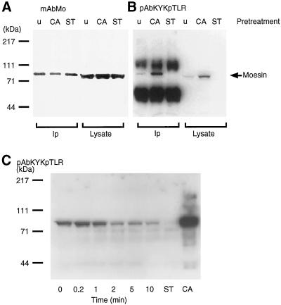
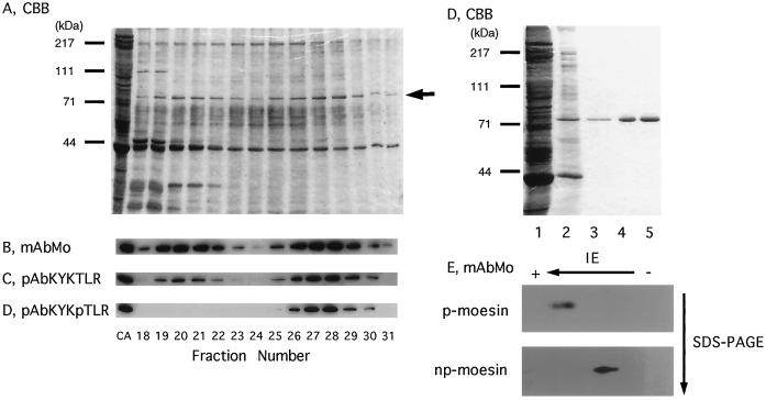
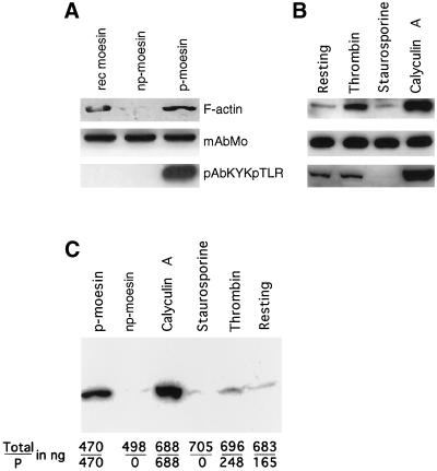
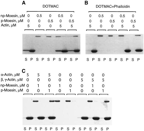
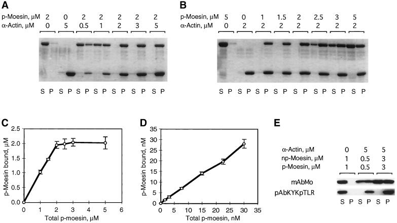
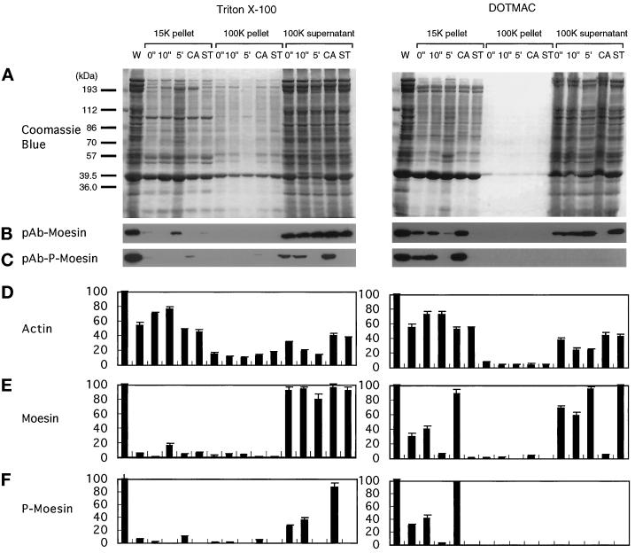
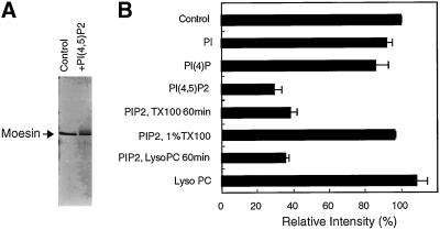
Activation of human platelets with thrombin transiently increases phosphorylation at (558)threonine of moesin as determined with phosphorylation state-specific antibodies. This specific modification is completely inhibited by the kinase inhibitor staurosporine and maximally promoted by the phosphatase inhibitor calyculin A, making it possible to purify the two forms of moesin to homogeneity. Blot overlay assays with F-actin probes labeled with either [32P]ATP or 125I show that only phosphorylated moesin interacts with F-actin in total platelet lysates, in moesin antibody immunoprecipitates, and when purified. In the absence of detergents, both forms of the isolated protein are aggregated. Phosphorylated, purified moesin co-sediments with alpha- or beta/gamma-actin filaments in cationic, but not in anionic, nonionic, or amphoteric detergents. The interaction affinity is high (Kd, approximately 1.5 nM), and the maximal moesin:actin stoichiometry is 1:1. This interaction is also observed in platelets extracted with cationic but not with nonionic detergents. In 0.1% Triton X-100, F-actin interacts with phosphorylated moesin only in the presence of polyphosphatidylinositides. Thus, both polyphosphatidylinositides and phosphorylation can activate moesin's high-affinity F-actin binding site in vitro. Dual regulation by both mechanisms may be important for proper cellular control of moesin-mediated linkages between the actin cytoskeleton and the plasma membrane.


Figures

Figure 1
Figure 1
Detection and quantification of moesin phosphorylation at a single site, 558threonine, in platelets by immunoblot analysis. (A) Immunoblot analysis with mouse monoclonal antibody (mAbMo) 38/87 of moesin immunoprecipitated with a polyclonal affinity purified anti-moesin reagent (pAbMo) from detergent extracts (Ip) and total platelet lysates: untreated (u), 100 nM calyculin A (CA), or 1 μM staurosporine (ST) pretreated for 10 min. (B) Immunoblot analysis with rabbit polyclonal anti-558T-p-moesin raised to the peptide (pAbKYKpTLR) of the same samples as in A. The position of moesin is indicated with an arrow. The amounts of moesin in each of the three lanes of Ip and lysates are approximately the same (A). Note in B the increase in phosphorylation in the CA samples and the absence of a phosphorylated band in the ST samples in comparison with untreated platelets. The two dense bands in the Ip lanes of B are heavy- and light-chain polypeptides of rabbit IgG in the immunoprecipitate. (C) Immunoblot analysis of moesin phosphorylation in response to thrombin stimulation of platelets. Aliquots of total platelet lysates were electrophoresed on 9% SDS-polyacrylamide gels and blotted after protein transfer to nitrocellulose with pAbKYKpTLR antibodies as in B. ST and CA refer to lysates from platelets pre-treated with staurosporine or calyculin A as in A. Molecular mass standards are shown on the left. The blot is representative of three separate experiments.
Figure 2
Figure 2
Purification of 558T-phosphorylated and unphosphorylated moesin from human platelets. Human platelets were treated with 100 nM calyculin A for 30 s at 37°C, which produces ∼50% of phosphorylated moesin (p-moesin). A cell lysate was prepared as described in MATERIALS AND METHODS and loaded onto a heparin-agarose column (15 × 57 mm). Proteins were eluted with a 200-ml linear gradient of 0–500 mM NaCl in equilibrating buffer at a flow rate of 1 ml/min, and fractions of 4 ml were collected and analysed by SDS-PAGE (A; CBB, Coomassie brilliant blue–stained gel) and immunoblot analysis (B–D). Analysis of fraction numbers 18–31 is shown, and a lysate of calyculin A-treated platelets served as the positive control in lane 1 (CA). Blots were developed with monoclonal moesin antibodies (B; mAbMo) and affinity-purified polyclonal moesin peptide (C; pAbKYKTLR) or moesin phosphopeptide antibodies (D; pAbKYKpTLR). The position of moesin is indicated by the arrow in A. In E, the Coomassie brilliant blue–stained gel shows the protein profile of fractions containing 558T-p-moesin in the different chromatographic steps: lane 1, total platelet lysate; lane 2, 558T-p-moesin eluted from heparin-agarose; lane 3, eluate from blue-Sepharose; lane 4, eluate from phenyl-Sepharose; and lane 5, final product after DEAE-cellulose. Molecular mass standards are shown in A and D. In F, immunoblots of purified 558T-p-moesin and nonphosphorylated (np-) moesin after two-dimensional electrophoretic separation show homogeneous spots. The blots were developed with monoclonal antibodies to moesin (mAbMo).
Figure 3
Figure 3
F-actin blot overlay detects binding to the phosphorylated form of moesin in platelet lysates and after purification. Proteins were separated by SDS-PAGE, transferred to nitrocellulose filters, and blotted with a 125I-labeled F-actin probe in A and B and with a [32P]ATP-labeled probe in C. Before this analysis, the amounts of immunoreactive moesin were estimated by densitometric analysis and comparison with known amounts of recombinant moesin. The loaded amount of moesin is indicated in nanograms. (A) Binding of F-actin to purified recombinant moesin made in bacteria (470 ng; rec moesin, lane 1) and to purified phosphorylated platelet moesin (500 ng; p-moesin, lane 3). Nonphosphorylated moesin (498 ng; np-moesin, lane 2) does not bind appreciably bind to the probe. Parallel blots were processed with monoclonal moesin antibodies (mAbMo) or with antibodies specific for phosphorylated moesin (pAbKYKpTLR), indicating comparable protein loads and lack of detectable phosphorylation signals for rec moesin and np-moesin. (B) F-actin and antibody blots of total lysates of resting platelets (lane 1; 496 ng), platelets activated with thrombin for 10 s (lane 2; 502 ng), and platelets treated for 10 min with staurosporine (483 ng) or calyculin A (497 ng). Note the increase in F-actin signal intensity caused by thrombin stimulation in comparison with resting platelets and in relation to the maximal signal detected after calyculin A pretreatment. This relationship is also apparent from the immunoblot with phosphorylation-specific antibodies. (C) The amount of purified phosphorylated (p-) and nonphosphorylated (np-) moesin and of the two forms of moesin in platelet lysates of calyculin A-, staurosporine-, and thrombin-treated or resting platelets was quantified by immunoblotting and scanning densitometry as described in MATERIALS AND METHODS. The data shown represent total moesin and p-moesin in nanograms (total/P). Note again the absence of F-actin binding to platelet moesin in the absence of phosphorylation and increased binding signal resulting from the increase in phosphorylation caused by thrombin stimulation or by calyculin A.
Figure 4
Figure 4
Phosphorylated moesin selectively co-sediments with F-actin. (A) Purified phosphorylated (p-) or nonphosphorylated (np-) platelet moesin (0.5 μM) was incubated either alone or together with rabbit skeletal α-F-actin (5 μM) in DOTMAC-containing buffer for 1 h at 25°C before centrifugation, as described in MATERIALS AND METHODS. Equal volumes of supernatant (S) and pellet (P) fractions were analysed by SDS-PAGE, and proteins were visualized by Coomassie brilliant blue staining. The top band corresponds to moesin; the bottom band is actin. In the absence of actin, all of the moesin remains in the supernatant. Mixed with F-actin, a large fraction of p-moesin, but only a small fraction of np-moesin, co-sediments and appears in the pellet fraction. As can be seen, a fraction of actin remains in the supernatant, presumably because of a DOTMAC-induced increase in critical concentration for actin polymerization. When phalloidin is added to stabilize F-actin, essentially all of the actin is pelleted; p-moesin, but not np-moesin co-sediments with these phalloidin-stabilized filaments (B). (C) Under identical co-sedimentation conditions, p-moesin sediments equally well with skeletal muscle α- and platelet β, γ-actin.
Figure 5
Figure 5
Phosphorylated moesin binds to filamentous actin with high affinity and at a ratio of 1:1. To define dissociation constants for the specific interaction between actin filaments and p-moesin in DOTMAC, co-sedimentation was done by keeping the p-moesin concentration fixed at 2 μM and varying the actin concentration from 0.5 to 5 μM (A) and by varying the p-moesin concentration from 1–5 μM at a fixed 2 μM concentration of actin (B). (C) Saturation binding analysis of p-moesin and α-actin at the micromolar level. Data were obtained under conditions described in B and are the mean ± SD of three separate measurements. p-Moesin was varied over the range shown, whereas actin concentration was constant at 2 μM. (D) Binding analysis of p-moesin and α-actin at the nanomolar level. p-Moesin was varied over the range shown, whereas actin concentration was constant at 200 nM. Data are the mean ± SD of three separate measurements derived from immunoblots probed with moesin antibodies. (E) Indicated amounts of p- and np-moesin were mixed before addition to actin filaments and centrifugation. The Western blot, developed with monoclonal antibodies to moesin (mAbMo) and p-moesin antibodies (pKYKpTLR), shows the distribution of the two moesin forms in supernatants (S) and pellets (P). Note that both forms remain in the supernatant in the absence of F-actin; all detectable p-moesin co-sediments with F-actin; increasing the amount of p- and np-moesin in the mixture yields expected amounts of p-moesin in the pellets, and np-moesin does not sediment as a mixed aggregate of p- and np-moesin.
Figure 6
Figure 6
Phosphorylation-dependent association of moesin with the DOTMAC-insoluble cytoskeleton in thrombin-activated platelets. Platelets were lysed with Triton X-100 (left panels) or DOTMAC (right panels) lysis buffer at the indicated times (0 and 10 s and 5 min) after stimulation with 1 NIH unit/ml human thrombin at 37°C. Platelets were also incubated with 100 nM calyculin A (CA) or 1 μM staurosporine (ST) for 10 min before thrombin activation for 10 min. Lysates were centrifuged for 4 min at 15,600 × g, and the first supernatants were centrifuged for a further 30 min at 100,000 × g. Low- and high-speed pellets and high-speed supernatants were solubilized in SDS sample buffer, and the proteins were resolved by SDS-PAGE on a 9% gel and transferred to nitrocellulose membranes. (A) Coomassie brilliant blue–stained gels of the Triton X-100 (left) and DOTMAC (right) extraction and fractionation. After transfer of the proteins to nitrocellulose, blots were incubated with polyclonal antibodies to moesin (pAb-moesin; B) or phosphorylated moesin (pAb-p-moesin; C). Relative amounts of actin (D), moesin (E), and phosphorylated moesin (F) were obtained by densitometric analysis of the gels shown in A and the corresponding immunoblots of B and C. The blots shown in B and C are representative of three separate experiments. The data presented in D–E represent the means ± SD of three separate measurements. For detailed description and explanation, see text.
Figure 7
Figure 7
Phosphatidylinositol 4,5-biphosphate is required for F-actin binding of phosphorylated moesin in Triton X-100. Phosphorylated (p-) and non-phosphorylated (np-) moesin isolated from platelets was incubated alone or in the presence of α-actin for 1 h at 37°C before sedimentation. In A, no detergent was added, and sizable fractions (∼60%) of p- and np-moesin sedimented and were recovered in the pellet together with actin. There was no apparent difference between the two forms of moesin. In B, 0.1% Triton X-100 was added to the reaction mixtures. Both forms of moesin were soluble and did not sediment under this condition. In C and D, mixed micelles of 0.1% Triton X-100 and 0.01% phosphatidylinositol (PI) or phosphatidylinositol 4,5-biphosphate (PIP2) were added to the reaction mixtures. Approximately 50% of the total p-moesin co-sedimented together with actin in the presence of PIP2. In all experiments, equal volumes of supernatant (S) and pellet (P) fractions were analysed by SDS-PAGE and Coomassie blue staining.
Figure 8
Figure 8
Gel shift induced by binding of phosphatidylinositol 4,5-biphosphate to moesin on SDS polyacrylamide gels. Phosphorylated or nonphosphorylated moesin (0.5 μg) was incubated with sonicated PI, PI(4), PI(4,5)P2, or LysoPC (lysophosphatidylcholine) in 10 μl of buffer F (final concentration, 0.02%, wt/vol) for 1 h at 37°C. In some experiments, 0.1% Triton X-100 or LysoPC was added, and the incubation was continued for an additional 1 h. The solutions were then solubilized with an equal volume of 2× SDS sample buffer and run on a 9% polyacrylamide gel under reducing conditions. Proteins were stained with the silver method. (A) An example is shown for moesin and PI(4,5)P2. Note the lower intensity of the band in the normal migration position. (B) The reduction in moesin under various conditions has been quantified by densitometry. The graph shows this loss as an indication of the gel shift that occurred in the presence of PI(4,5)P2. This shift in migration is weakly influenced by a 60-min incubation in 0.1% Triton X-100 or LysoPC but is completely reversed in 1% Triton X-100.
Figure 9
Figure 9
A multistep model for the regulation of the F-actin-binding activity of moesin in platelets. In the resting state, moesin is bound to as-yet-unknown membrane proteins or sites indicated by phosphatidylinositol polyphosphate (PIPx). Membrane-bound moesin may exist in equilibrium with a soluble, cytosolic form. A first signal, perhaps involving PI(P) kinases, elevates levels of PIP2. Moesin in a complex with other membrane components undergoes a conformational change, allowing it to become a substrate for a membrane-associated kinase, such as PKC or Rho-kinase. After phosphorylation at 558Thr, the F-actin binding function of moesin is activated, and linkage with actin filaments in the vicinity of the membrane may occur. Steady-state phosphorylation is maintained by the action of protein kinase(s) and protein phosphatase(s). Staurosporine and calyculin A shift the balance toward complete dephosphorylation or phosphorylation, respectively. Similarly, transient changes in the number of phosphorylated and activated moesin molecules, caused by an increase in kinase or a decrease in phosphatase activity, enhance the potential to form new membrane–cytoskeletal links.

Similar articles

Cited by

References

    1. Algrain M, Turunen O, Vaheri A, Louvard D, Arpin M. Ezrin contains cytoskeleton and membrane binding domains accounting for its proposed role as a membrane-cytoskeletal linker. J Cell Biol. 1993;120:129–139. - PMC - PubMed
    1. Amieva MR, Furthmayr H. Subcellular localization of moesin in dynamic filopodia, retraction fibers, and other structures involved in substrate exploration, attachment, and cell-cell contacts. Exp Cell Res. 1995;219:180–196. - PubMed
    1. Amieva MR, Litman P, Huang L, Ichimaru E, Furthmayr H. Disruption of dynamic cell surface architecture of NIH3T3 fibroblasts by the N-terminal domains of moesin and ezrin: in vivo imaging with GFP fusion proteins. J Cell Sci. 1998;112:111–125. - PubMed
    1. Benka ML, et al. The thrombin receptor in human platelets is coupled to a GTP binding protein of the G alpha q family. FEBS Lett. 1995;363:49–52. - PubMed
    1. Bretscher A. Rapid phosphorylation and reorganization of ezrin and spectrin accompany morphological changes induced in A431 cells by epidermal growth factor. J Cell Biol. 1989;108:921–930. - PMC - PubMed

Publication types

MeSH terms