. 1999 Aug 15;19(16):6748-54.
doi: 10.1523/JNEUROSCI.19-16-06748.1999.

Distinct functional types of associative long-term potentiation in neocortical and hippocampal pyramidal neurons

Affiliations

Distinct functional types of associative long-term potentiation in neocortical and hippocampal pyramidal neurons

D V Buonomano. J Neurosci. .

Abstract

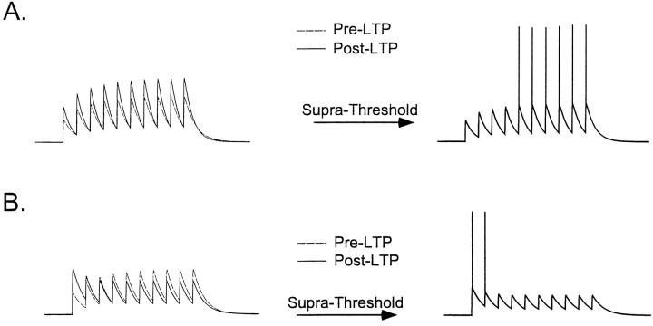
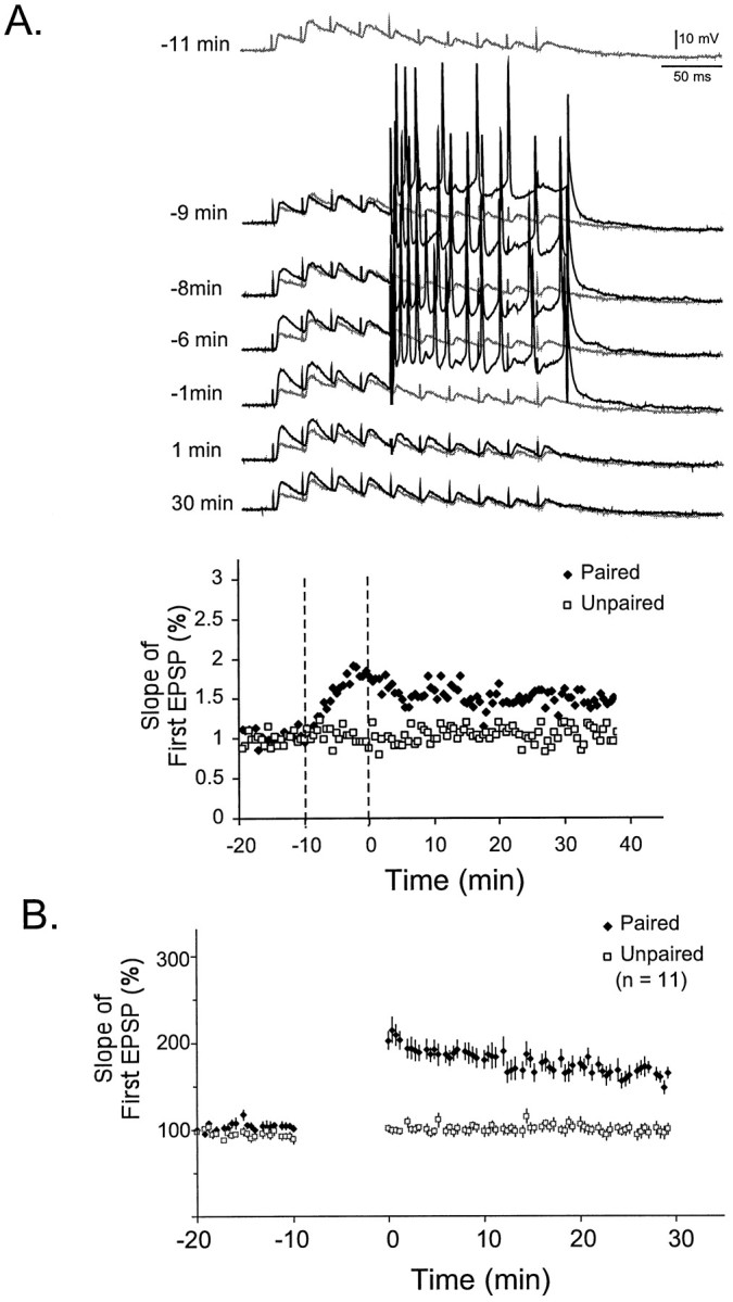
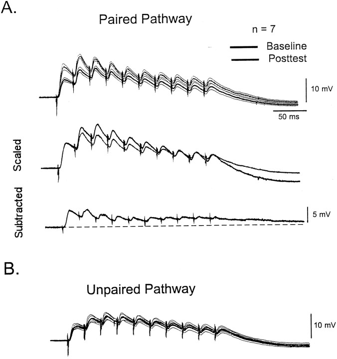
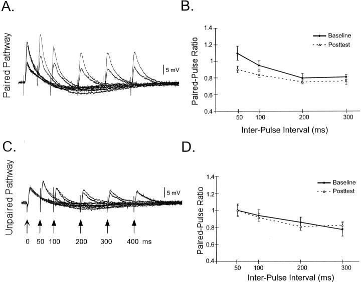
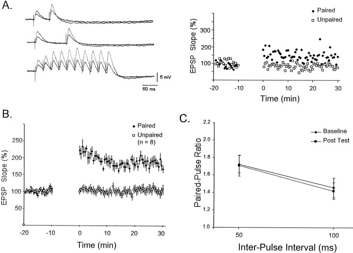
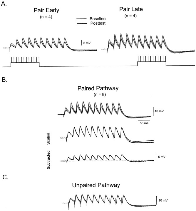
The response of a neuron to a time-varying stimulus is influenced by both short- and long-term synaptic plasticity. Both these forms of plasticity produce changes in synaptic efficacy of similar magnitude on very different time scales. A full understanding of the functional role of each form of plasticity relies on understanding how they interact. Here we examine how long-term potentiation (LTP) and short-term plasticity (STP) interact in two different cell types that exhibit NMDA-dependent LTP: neocortical L-II/III and hippocampal CA1 pyramidal cells. STP was examined using both paired pulses and trains of pulses before and after the induction of LTP. In both cell types, the same pairing protocol was used to induce LTP in the presence of an unpaired control pathway. Pairing produced a robust increase in the amplitude of the first EPSP both in the neocortex and hippocampus. However, although in CA1 neurons the same degree of potentiation was maintained throughout the duration of a brief stimulus train, in L-II/III neurons relatively less potentiation was seen in the later EPSPs of the train. Paired-pulse analyses revealed that a uniform potentiation is observed at intervals >100 msec, but at shorter intervals there is a preferential enhancement of the first pulse. Thus, in the cortex LTP may preferentially amplify stimulus onset. These results suggest that there are distinct forms of associative LTP and that the different forms may reflect the underlying computations taking place in different areas.


Figures

Fig. 1.
Fig. 1.
Schematic of how the interaction between LTP and STP can effect neuronal responses to temporal stimuli. If a cell initially exhibits some facilitation in response to a train of inputs, LTP can result in two different scenarios. A, If the induction of LTP does not modify the short-term facilitation, the temporal response characteristics remain unchanged. Suprathreshold responses will reflect the facilitation and produce a neuron that selectively responds to prolonged inputs. B, If LTP modifies short-term plasticity by producing proportionally larger potentiation of the first EPSP, suprathreshold responses will favor detection of the stimulus onset and not of the later temporal features of a stimulus.
Fig. 2.
Fig. 2.
Induction of LTP in a L-II/III pyramidal neuron.A, Example of the induction of LTP by pairing depolarization with presynaptic activity. The top traceshows the postsynaptic response to 10 pulses (40 Hz) before training (−11 min). Subsequent traces show the responses during and after induction of LTP. During the induction protocol, depolarization was paired with the latter half of the 40 Hz train. The baseline trace is repeated for comparison at each point (gray). Thebottom panel shows the slope of the first EPSP during the paired and unpaired pathway. B, Average LTP from 11 experiments in L-II/III pyramidal neurons.
Fig. 3.
Fig. 3.
Interaction of LTP and STP in L-II/III neurons.A, Each trace represents the average response of seven neurons to the 40 Hz train before (black) and after (gray) the induction of LTP. The thin lines represent the SEM of the average trace. The middle traces are the same as those displayed above after scaling the first EPSP of the baseline response to the first EPSP of the posttest response. Note that for the second to fifth EPSPs the baseline responses are larger, indicating relatively less potentiation of these EPSPs. The bottom trace shows the subtraction of the baseline from the posttest trace. B, Average postsynaptic responses to the unpaired pathway in the same seven cells.
Fig. 4.
Fig. 4.
LTP modifies paired-pulse plasticity in L-II/III neurons. Traces from a single experiment before (black) and after (gray) the induction of LTP in the paired (A) and unpaired (C) pathways. Right panels, Average paired-pulse ratio (slope of the second pulse/slope of first pulse) before and after the induction of LTP for the paired (B) and unpaired (D) pathways. In B, each point represents an average of 15, 15, 6, and 6 cells for the four different intervals (in ascending order). The values of 50 and 100 msec are significantly different before and after LTP (p < 0.02). InD, the number of cells for each point was 12, 12, 8, and 8. Note that in A the apparent facilitation at 50 msec is mostly temporal summation. The paired-pulse ratios shown inB and D are calculated from the slopes of the second EPSP after subtraction of the first EPSP.
Fig. 5.
Fig. 5.
A, LTP in hippocampal CA1 pyramidal neurons. Traces of a single experiment showing the responses to paired-pulse stimulation at intervals of 50 and 100 msec, and a 40 Hz train before (black) and after LTP (gray). Note that the differences in the amplitude of the first EPSP in the three different conditions reflect the normal variability of synaptic amplitude. The slope of the first EPSP is shown throughout the experiment for the paired and unpaired pathway on the right. B, Average LTP for all eight cells. C, Average paired-pulse ratio for the same eight cells. There was no significant change in the paired-pulse ratio after the induction of LTP for either the 50 or 100 msec interval.
Fig. 6.
Fig. 6.
In the hippocampus, LTP uniformly potentiates all EPSPs of a train. A, Two different protocols were used to induce LTP. Depolarization was paired either with the first five pulses (early) or last five pulses (late). Each trace shows the average of four separate experiments before (black) and after (gray) the induction of LTP. Thin lines represent the SEM. In both groups, potentiation was uniform across the length of the train. B,Top traces represent the average baseline and posttest trace from all eight cells. Middle traces are the same as those shown above after scaling. Note that the overlap of the normalized traces indicate potentiation was the same across all EPSPs.Bottom trace shows the subtraction of the baseline from the posttest traces. C, In the unpaired pathway there were no significant changes before and after the induction of LTP.

References

    1. Abbott LF, Varela JA, Kamal S, Nelson SB. Synaptic depression and cortical gain control. Science. 1997;275:220–224. - PubMed
    1. Armstrong-James M, Diamond M, Ebner FF. An innocuous bias in whisker use in adult rats modifies receptive fields of barrel cortex neurons. J Neurosci. 1994;14:6978–6991. - PMC - PubMed
    1. Buonomano DV, Merzenich MM. Temporal information transformed into a spatial code by a neural network with realistic properties. Science. 1995;267:1028–1030. - PubMed
    1. Buonomano DV, Merzenich MM. Associative synaptic plasticity in hippocampal CA1 neurons is not sensitive to unpaired presynaptic activity. J Neurophysiol. 1996;76:631–636. - PubMed
    1. Buonomano DV, Merzenich MM. Cortical plasticity: from synapses to maps. Annu Rev Neurosci. 1998a;21:149–186. - PubMed

Publication types

LinkOut - more resources