. 1999 Aug 15;19(16):6755-66.
doi: 10.1523/JNEUROSCI.19-16-06755.1999.

Active role of glutamate uptake in the synaptic transmission from retinal nonspiking neurons

Affiliations

Active role of glutamate uptake in the synaptic transmission from retinal nonspiking neurons

K Matsui et al. J Neurosci. .

Abstract

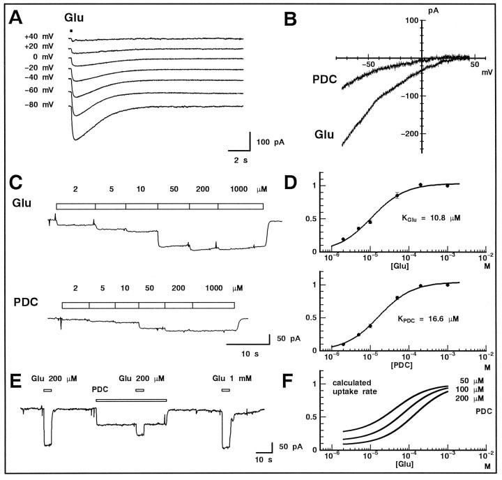
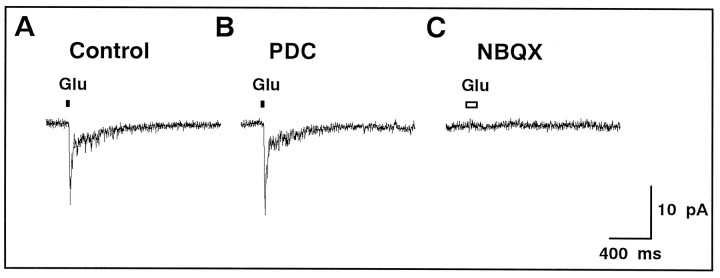
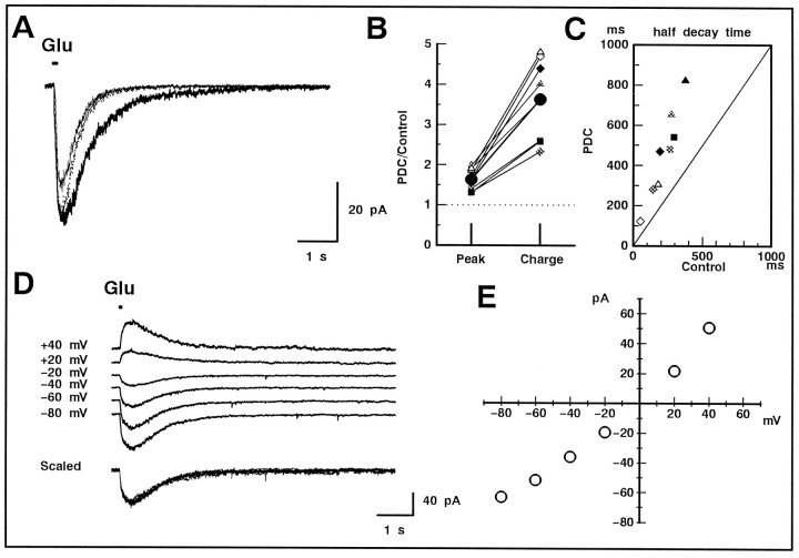
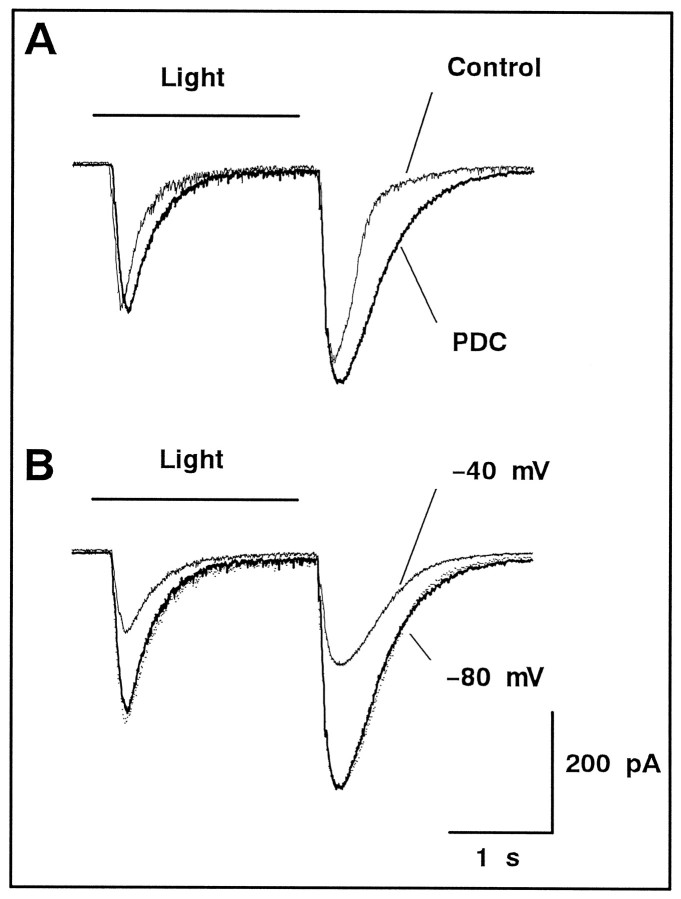
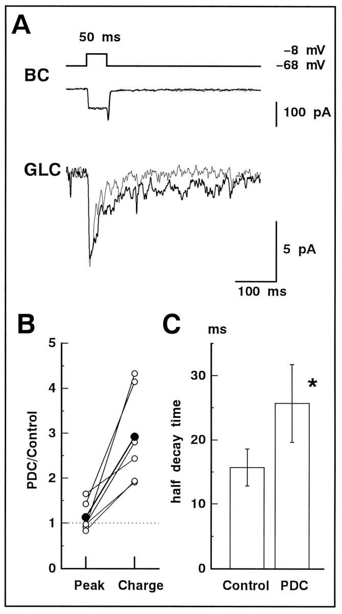
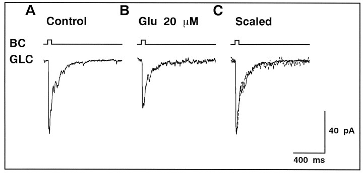
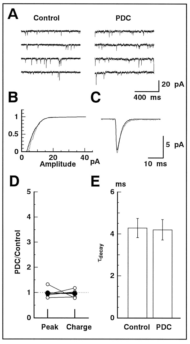
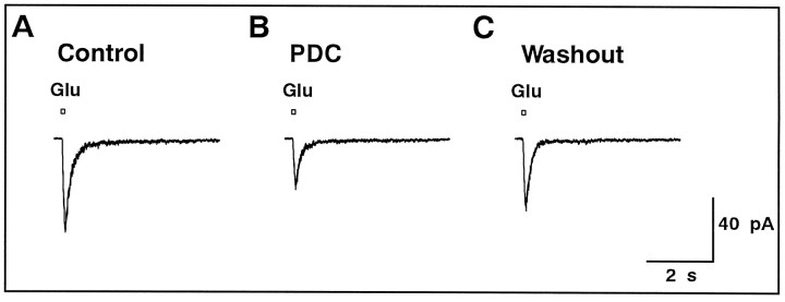
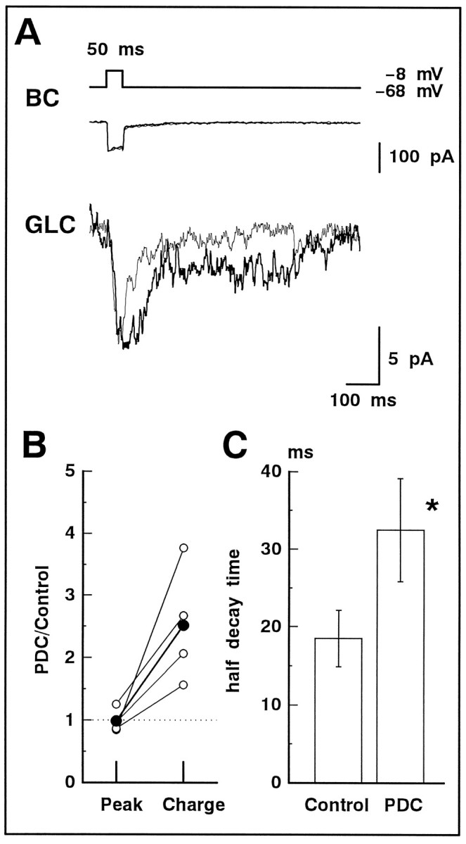
We examined the role of glutamate uptake in the synaptic transmission of graded responses from newt retinal bipolar cells (BCs) to ganglion layer cells (GLCs). In dissociated Müller cells (retinal glia), glutamate evoked an uptake current that was effectively inhibited by L-trans-pyrrolidine-2,4-dicarboxylic acid (PDC). PDC had no effect on the non-NMDA receptors of dissociated spiking neurons. In the retinal slice preparation, dual whole-cell recordings were performed from a pair of BC and GLC. A depolarizing pulse applied to a BC activated the Ca(2+) current (I(Ca)) in the BC and evoked an EPSC in the GLC. Application of PDC prolonged both non-NMDA and NMDA receptor-mediated components of the evoked EPSC but changed neither the amplitude nor time course of I(Ca). When the slice preparation was superfused with a solution containing glutamate but not PDC, the evoked EPSC decreased in amplitude without changing the time course, suggesting that the prolongation of the evoked EPSC is not attributable to a simple increase in ambient glutamate concentration after inhibition of glutamate uptake. Because PDC did not affect the amplitude, time course, or frequency of spontaneous EPSCs, it is unlikely that PDC modified presynaptic and/or postsynaptic mechanisms. These results indicate that inhibition of glutamate uptake slows the clearance of glutamate accumulated in the synaptic cleft by multiple quantal release and prolongs the evoked EPSC. The role of glutamate uptake at synapses in the inner retina is not only to maintain the extracellular glutamate concentration at a low level but also to terminate the light-evoked EPSCs rapidly.


Figures

Fig. 1.
Fig. 1.
PDC blocked glutamate uptake in Müller cells isolated from the newt retina. A, Glutamate (1 mm) was puff applied (100 msec; timing indicated as thebar at the top) to a Müller cell that was whole-cell voltage-clamped at various potentials (noted on theleft). The glutamate-induced uptake current did not reverse its polarity at positive potentials. B,I–V curves obtained in the presence of 1 mmglutamate (Glu) and 200 μm PDC. Membrane currents were measured by applying voltage ramps (130 mV/300 msec). Each I–V curve was derived from the difference between the average of three current traces in the presence and absence of the chemicals, which were applied via the Y-tube microflow system.C, The uptake currents induced by various concentrations (top) of glutamate (Glu) or PDC. Bothtraces were obtained from the same cell voltage-clamped at −75 mV. D, Dose–response curves for glutamate (top panel) or PDC (bottom panel). Means ± SEM are illustrated (pooled data from 16 cells). Data points were fitted by the Michaelis–Menten equation. E, Inhibition of the glutamate-induced uptake current by PDC (200 μm). The concentration of glutamate (Glu) was 200 μm (left andmiddle) and 1 mm (right). The cell was held at −75 mV. F, Estimation of the uptake rate of glutamate in the presence of PDC. A simple competitive inhibition model was used with the values ofKGlu and KPDCobtained in D. The concentration of PDC is shown at theright of the curves.
Fig. 2.
Fig. 2.
The current through non-NMDA receptors was not affected by PDC. A, A spiking neuron isolated from the newt retina was voltage-clamped at −80 mV. Both the superfusate and the puff pipette solution included 50 μmd-AP-5 to block the current through NMDA receptors. A 20 msec puff-application (top) of 200 μmglutamate (Glu) induced an inward current.B, The glutamate-induced current was not affected by the presence of 200 μm PDC. C, Application of NBQX (5 μm) to the superfusate completely blocked the response to a 100 msec puff of glutamate.
Fig. 3.
Fig. 3.
Effect of PDC on the non-NMDA receptor-mediated current in GLCs of the retinal slice. A, A GLC was voltage-clamped at −80 mV, and glutamate (200 μm) was puff applied for 100 msec. The tip of the puff pipette was carefully positioned above the IPL to evoke a large response. In both the bath and puff solutions, divalent cations were replaced with Co2+ to suppress the synaptic transmission, andd-AP-5 (50 μm) was included to block the NMDA receptor-mediated current. The glutamate-induced current in the presence of 200 μm PDC (thick trace) was larger in amplitude and decayed more slowly than control (thin trace). After scaling (dotted trace), it is clear that PDC prolonged the glutamate-induced current through non-NMDA receptors. B, Relative increase in the peak amplitude (Peak) and total charge (Charge) before and after the application of PDC. Data were obtained from three cells and are illustrated with different symbols. Duration of the puff was 10 (open), 50 (half-tone), or 100 (filled) msec. The averaged values are shown with large filled circles. C, The scatter diagram illustrates the relationship between the half-decay time of the glutamate-induced current in the absence (Control) and presence of PDC. Allsymbols correspond to those shown in B. D, The current was evoked in a GLC by a 50 msec puff of glutamate in the presence of 200 μm PDC. The holding potential was changed to various values. The current traces are shifted arbitrarily for a better view. All of the current traces were superimposable after scaling (bottom traces).E, The peak amplitude of the glutamate-induced current was plotted against the command potential of GLC. The linear relationship confirmed that the glutamate-induced current was caused by the activation of non-NMDA receptors.
Fig. 4.
Fig. 4.
The light-evoked responses in GLC were enhanced by PDC. A, With full-field illumination (Light), an ON/OFF transient response (thin trace) was evoked in GLC of the retinal slice, which was superfused with the solution containing d-AP-5 (50 μm). Both ON and OFF transients were prolonged by application of 200 μm PDC (thick trace). GLC was held at −80 mV. B, The photoresponses were recorded in the presence of PDC. The holding potential was set at −80 (thick trace) and −40 (thin trace) mV. After scaling (dotted trace), both traces were superimposable, indicating that the NMDA receptor-mediated current was not emerged by application of PDC.
Fig. 5.
Fig. 5.
The evoked non-NMDA-EPSC was prolonged by PDC. A, A 50 msec depolarizing pulse (from −68 to −8 mV; top) applied to BC activatedICa (middle; thin trace) in BC and evoked non-NMDA-EPSC in GLC voltage-clamped at −80 mV (bottom; thin trace). After application of 200 μm PDC, neither the amplitude nor time course of ICa(thick trace) was affected, but the decay of the non-NMDA-EPSC was significantly prolonged (thick trace). d-AP-5 (50 μm) was included in the bath solution. The current traces of BC for this and the subsequent figures are shown after leak subtraction. B,Relative increase in the peak amplitude (Peak) and total charge (Charge) of the evoked non-NMDA-EPSC before and after application of PDC. Data were obtained from six cell pairs. The averaged values are shown bylarge filled circles. C, The half-decay time of the evoked non-NMDA-EPSC in the absence (Control) and presence of PDC. Asterisks in this and subsequent figures indicate that the difference is statistically significant (p< 0.05). Data were obtained from six cell pairs.
Fig. 6.
Fig. 6.
Elevation of the ambient glutamate concentration did not prolong the evoked non-NMDA-EPSC.A, BC was depolarized from −68 to −8 mV for 50 msec, and the evoked non-NMDA-EPSC was recorded from GLC voltage-clamped at −80 mV. Activation of NMDA receptors was blocked by 50 μmd-AP-5 in the bath solution.B, Addition of 20 μm glutamate to the bath solution reduced the amplitude of the evoked non-NMDA-EPSC. C, After scaling of the trace shown in B (dotted trace), both current traces were superimposable.
Fig. 7.
Fig. 7.
Spontaneous EPSCs were not affected by PDC.A, The membrane current was recorded continuously (4 sec records are displayed) from a GLC voltage-clamped at −80 mV. Spontaneous EPSCs were clearly observed. The slice preparation was superfused with the solution containing d-AP-5 (50 μm; Control) and then with the solution containing d-AP-5 and PDC (200 μm).B, The cumulative amplitude distribution of spontaneous EPSCs obtained in the absence (thick line) and presence (thin line) of PDC. C, The waveforms of the averaged spontaneous EPSCs in the absence (thick line) and presence (thin line) of PDC.D, Relative changes of the peak amplitude and total charge of the averaged spontaneous EPSCs in the absence and presence of PDC (n = 5). The averaged values of five data are shown by large filled circles. E, The decay phase of the averaged spontaneous EPSC was well fitted by a single exponential function. The time constant of the decay (τdecay) was not affected by PDC. Data were obtained from five cells.
Fig. 8.
Fig. 8.
Effect of PDC on the NMDA receptor-mediated current in an isolated spiking neuron. A, A 100 msec puff (top) of glutamate (200 μm) evoked an inward current in an isolated spiking neuron voltage-clamped at −40 mV. Both the superfusate and the puff pipette solution included 5 μm NBQX, 10 μm glycine, and 10 μm strychnine. B, Bath application of PDC (200 μm) reduced the amplitude of the glutamate-induced current. C, The suppressive effect of PDC reversed after washout.
Fig. 9.
Fig. 9.
The evoked NMDA-EPSC was prolonged by PDC. A, In the slice preparation, dual whole-cell recordings were performed. A 50 msec depolarizing pulse (from −68 to −8 mV; top) applied to BC activatedICa (middle; thin trace) in BC and evoked the NMDA-EPSC in GLC voltage-clamped at −40 mV (bottom; thin trace). Bath application of 200 μm PDC did not change ICa (middle;thick trace) but prolonged the evoked NMDA-EPSC (bottom; thick trace). The superfusate always contained 5 μmNBQX. B, Relative increase in the peak amplitude (Peak) and total charge (Charge) of the evoked NMDA-EPSC before (Control) and after application of PDC (n = 4). The averaged values are shown by large filled circles. C, The half-decay time of the evoked NMDA-EPSC in the absence (Control) and presence of PDC.

References

    1. Ball AK, Dickson DH. Displaced amacrine and ganglion cells in the newt retina. Exp Eye Res. 1983;36:199–213. - PubMed
    1. Barbour B, Brew H, Attwell D. Electrogenic uptake of glutamate and aspartate into glial cells isolated from the salamander (Ambystoma) retina. J Physiol (Lond) 1991;436:169–193. - PMC - PubMed
    1. Brew H, Attwell D. Electrogenic glutamate uptake is a major current carrier in the membrane of axolotl retinal glial cells. Nature. 1987;327:707–709. - PubMed
    1. Bridges RJ, Stanley MS, Anderson MW, Cotman CW, Chamberlin AR. Conformationally defined neurotransmitter analogues. Selective inhibition of glutamate uptake by one pyrrolidine-2:4-dicarboxylate diastereomer. J Med Chem. 1991;34:717–725. - PubMed
    1. Clements JD. Transmitter timecourse in the synaptic cleft: its role in central synaptic function. Trends Neurosci. 1996;19:163–171. - PubMed

Publication types

MeSH terms

LinkOut - more resources