

doi: 10.1523/JNEUROSCI.19-16-06907.1999.
Embryonic lethal abnormal vision-like RNA-binding proteins regulate neurite outgrowth and tau expression in PC12 cells
Affiliations
- PMID: 10436048
- PMCID: PMC6782881
- DOI: 10.1523/JNEUROSCI.19-16-06907.1999
Item in Clipboard
Embryonic lethal abnormal vision-like RNA-binding proteins regulate neurite outgrowth and tau expression in PC12 cells
J Neurosci.
.
Abstract
The embryonic lethal abnormal vision (ELAV)-like proteins are mRNA-binding proteins that regulate mRNA stability. The neuronal members of this family are required for neuronal differentiation. We identified the binding region of purified HuD protein to a target neuronal mRNA encoding for the tau microtubule-associated protein and demonstrated an in vivo interaction between the ELAV-like protein and its target tau mRNA. We show that treatment of neuronal cells with antisense oligodeoxynucleotides directed against HuD blocks the induction of neurite outgrowth and decreases the levels of tau mRNAs, indicating that the ELAV-like proteins are required for neuronal differentiation.
Figures

Effect of r-HuD antisense treatment on neurite outgrowth in PC12 cells. A, Control PC12 cells treated with NGF for 4 d. B, PC12 cells treated with NGF and r-HuD antisense oligonucleotides for 4 d. C, PC12 cells treated with NGF and unrelated AC6 antisense oligonucleotides. D, PC12 cells treated with NGF and sense r-HuD oligonucleotides for 4 d. Scale bar, 20 μm.
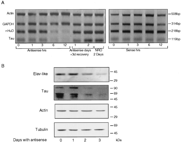
Effect of r-HuD antisense treatment on tau and r-HuD levels in PC12 cells. A, RT-PCR of RNA isolated from PC12 cells treated with NGF and r-HuD antisense for the specified times (hrs) and from cells allowed to recover for 3 d after 1 or 2 d of r-HuD antisense treatment. NROindicates RNA isolated from PC12 cells treated for 2 d with NGF and unrelated AC6 antisense oligos. Sense panel shows RT-PCR results for PC12 cells treated with NGF and sense HuD oligo for the specified times (hrs). RT-PCR was performed using r-HuD, tau, actin, and GAPDH primers in each sample. B, Immunoblot analysis of ELAV-like, tau, actin, and tubulin proteins in cell extracts prepared from PC12 cells treated for 3 d with NGF and r-HuD antisense oligos.
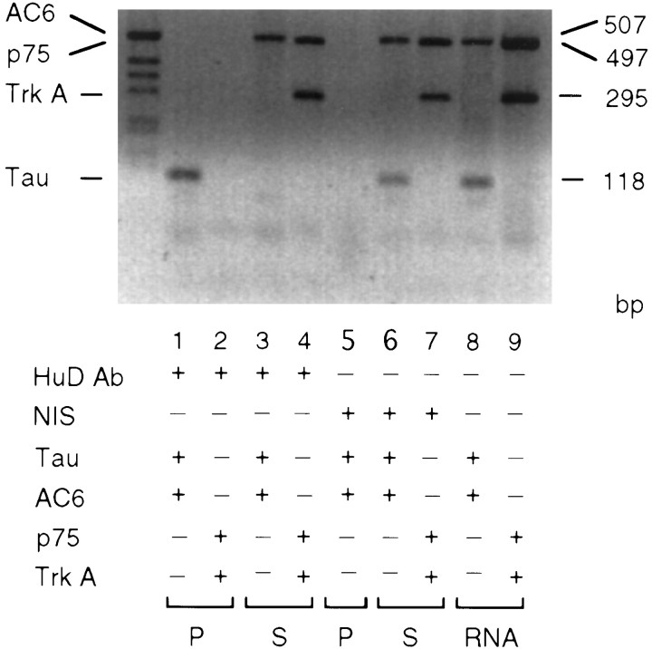
Binding of tau mRNA to ELAV-like proteinsin vivo: immunoprecipitation of tau mRNA–ELAV-like complex, followed by isolation and RT-PCR analysis of RNA. PC12 cell extracts were immunoprecipitated with anti-Hu serum. The RNA isolated from the immunoprecipitated complex (P) and the remaining supernatant (S) were assayed by RT-PCR with tau and AC6 primers (lanes 1, 3) and with p75NGFR and TrkA primers (lanes 2, 4). Lanes 5–7, Cell extracts immunoprecipitated with nonimmune serum (NIS) were assayed with tau and AC6-specific primers (lane 5, 6) and with p75NGFR and TrkA primers (lane 7). RT-PCR products using total RNA isolated from PC12 cells were amplified with tau and AC6-specific primers (lane 8) and with p75NGFR and TrkA primers (lane 9). The sizes of RT-PCR products obtained with AC6, p75NGFR, TrkA, and tau primers are 507, 497, 295 and 118 bp, respectively.
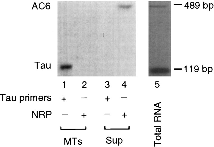
Association of tau mRNA-ELAV-like protein complex with MTs in vivo. Preexisting polymerized microtubules (MTs) and unpolymerized supernatant (Sup) fractions were prepared from PC12 cells. The MT fraction was immunoprecipitated by anti-Hu serum. RNA was isolated from the MT-immunoprecipitated complex and the initial supernatant, and analyzed by RT-PCR using tau-specific (lanes 1, 3) and AC6-specific NRP (lanes 2, 4) primers.Lane 5 is similar to lane 8 in Figure3.
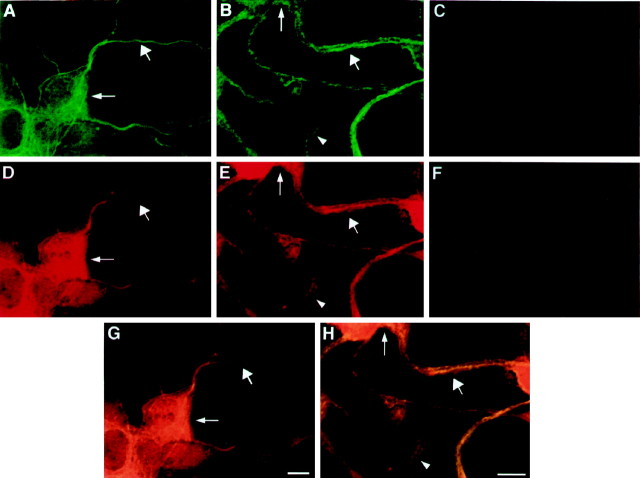
Confocal microscopy analysis of PC12 cells stained with tubulin and anti-ELAV-like antibodies. A,B, Confocal image of PC12 cells stained with tubulin antibodies. D, E, Confocal image of PC12 cells stained with anti-Hu serum. C, F, Control experiments showing no penetration of Cy3 or fluorescein signals into the opposite windows. Thin arrow,wide arrow, and arrowhead indicate cell body, neurite, and growth cone, respectively. Magnification: 5× forA, D, G; 6× forB, E, H. Scale bar, 5 μm.

Structure of tau mRNA. The fragments (F, G, H,I, and J) used in this study correspond to nucleotides 1778–2175 (397 bp), 2175–2760 (624 bp), 2519–2760 (241 bp), 2519–2610 (91 bp), and 2610–2760 (150 bp), respectively (Sadot et al., 1994). The 241-nucleotide sequence of fragment H is presented, and the I fragment containing the U-rich segment is boxed. The U-rich 21 nucleotides are deleted from fragment G (Gdel).

Immunoprecipitation of UV cross-linked complexes by anti-Hu ELAV-like antibodies: SDS gel analysis of immunoprecipitated complexes formed between PC12 cell extracts (lane 1), assembled MT preparations isolated from PC12 cells (lane 2), or purified HuD-GST (lane 3) UV cross-linked to [32P]-labeled tau RNA fragment I. NIS is the complex formed with normal serum (lane 4).Lanes 5 and 6 show unprecipitated PC12 extract and GST-HuD protein analyzed immediately after cross-linking.
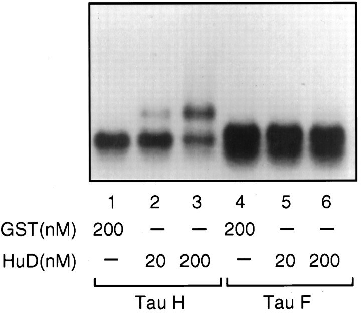
Purified HuD binds to tau mRNA. The indicated [32P]-labeled RNAs were incubated with the indicated concentrations of GST or HuD. After incubation, the reaction mixture was resolved by gel electrophoresis in 0.8% agarose gel.
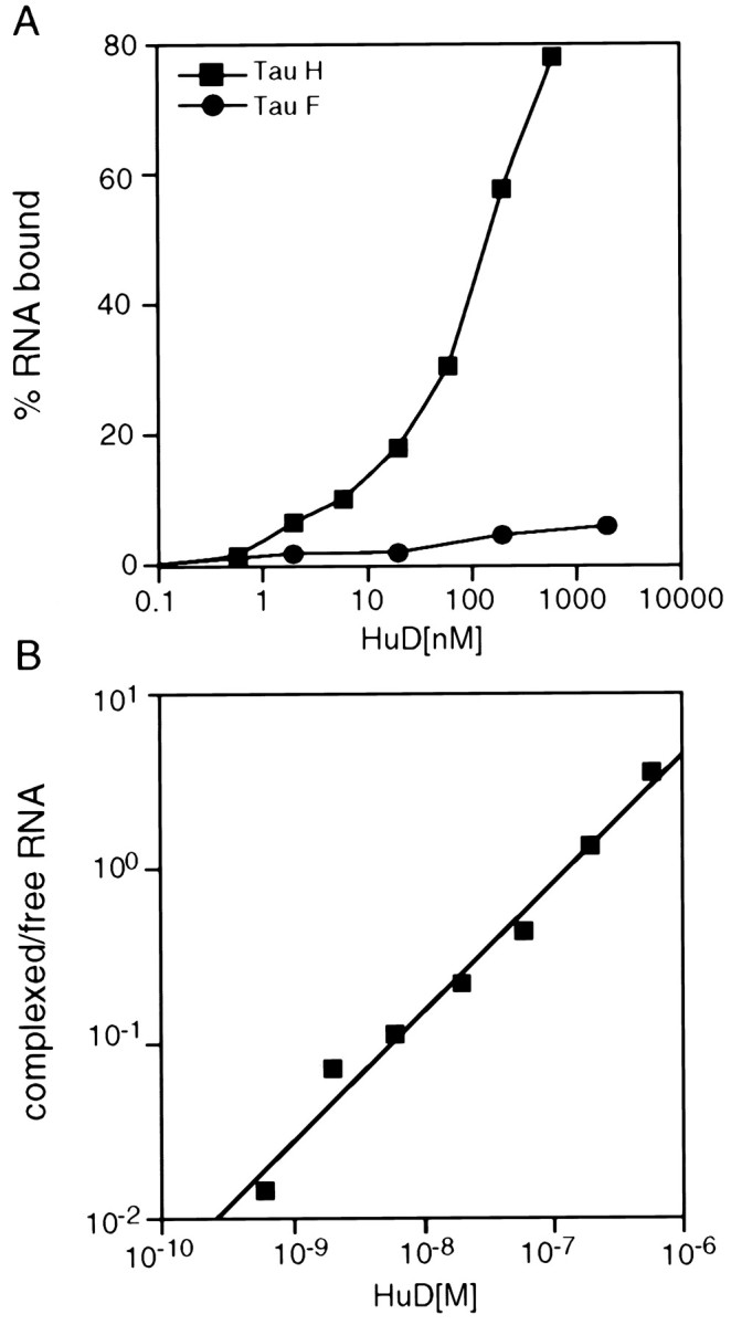
Affinity of HuD for tau mRNA. The affinity of purified HuD-GST for tau mRNA was determined by nitrocellulose filter binding assay, as described in Materials and Methods. A, Plot of percentage of bound RNA versus log of HuD concentration.B, Plot of log of ratio between complexed/free RNA versus log of HuD concentration.
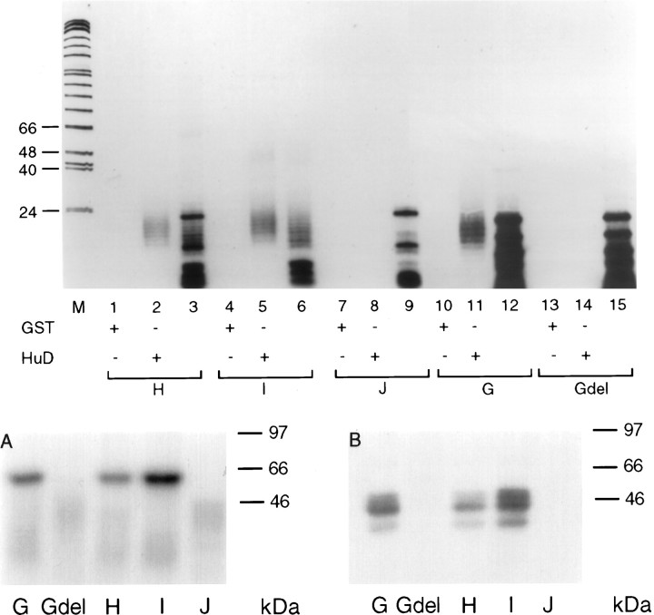
RNase T1 selection analysis of the HuD binding region in the tau-H fragment. RNase T1 selection assay was performed as described previously (Chung et al., 1996) with GST (lanes 1, 4, 7, 10, 13) or HuD (lanes 2, 5, 8, 11, 14), incubated with [32P]-labeledH, I, J, G, and Gdel RNAs, respectively. The reaction mixture was treated with RNase T1 and filtered through nitrocellulose to select for the RNA/protein complexes. After washing, the fragments were eluted and resolved on a 12% denaturing polyacrylamide gel. Lanes 3, 6, 9, 12, and 15 are the total unselected RNase T1 digests of the indicated fragments. A, UV cross-linking assay with purified GST-HUD. B, UV cross-linking assay with PC12 protein extracts.
Similar articles
-
The RNA-binding protein HuD is required for GAP-43 mRNA stability, GAP-43 gene expression, and PKC-dependent neurite outgrowth in PC12 cells.Mol Biol Cell. 2000 Sep;11(9):3191-203. doi: 10.1091/mbc.11.9.3191. Mol Biol Cell. 2000. PMID: 10982410 Free PMC article.
-
Cytoplasmic localization is required for the mammalian ELAV-like protein HuD to induce neuronal differentiation.Genes Cells. 1999 Nov;4(11):667-83. doi: 10.1046/j.1365-2443.1999.00292.x. Genes Cells. 1999. PMID: 10620013
-
Overexpression of HuD, but not of its truncated form HuD I+II, promotes GAP-43 gene expression and neurite outgrowth in PC12 cells in the absence of nerve growth factor.J Neurochem. 2000 Sep;75(3):1103-14. doi: 10.1046/j.1471-4159.2000.0751103.x. J Neurochem. 2000. PMID: 10936192
-
Role of HuD and other RNA-binding proteins in neural development and plasticity.J Neurosci Res. 2002 Apr 15;68(2):121-6. doi: 10.1002/jnr.10175. J Neurosci Res. 2002. PMID: 11948657 Review.
-
Emerging complexity of the HuD/ELAVl4 gene; implications for neuronal development, function, and dysfunction.RNA. 2013 Aug;19(8):1019-37. doi: 10.1261/rna.039164.113. RNA. 2013. PMID: 23861535 Free PMC article. Review.
Cited by
-
Neuron-specific ELAV/Hu proteins suppress HuR mRNA during neuronal differentiation by alternative polyadenylation.Nucleic Acids Res. 2012 Mar;40(6):2734-46. doi: 10.1093/nar/gkr1114. Epub 2011 Dec 1. Nucleic Acids Res. 2012. PMID: 22139917 Free PMC article.
-
The RNA-binding protein HuD is required for GAP-43 mRNA stability, GAP-43 gene expression, and PKC-dependent neurite outgrowth in PC12 cells.Mol Biol Cell. 2000 Sep;11(9):3191-203. doi: 10.1091/mbc.11.9.3191. Mol Biol Cell. 2000. PMID: 10982410 Free PMC article.
-
Association between the neuron-specific RNA-binding protein ELAVL4 and Parkinson disease.Hum Genet. 2005 Jun;117(1):27-33. doi: 10.1007/s00439-005-1259-2. Epub 2005 Apr 13. Hum Genet. 2005. PMID: 15827745
-
MRNA stability and the control of gene expression: implications for human disease.Neurochem Res. 2002 Oct;27(10):957-80. doi: 10.1023/a:1020992418511. Neurochem Res. 2002. PMID: 12462398 Review.
-
Post-transcriptional regulatory elements and spatiotemporal specification of neocortical stem cells and projection neurons.Neuroscience. 2013 Sep 17;248:499-528. doi: 10.1016/j.neuroscience.2013.05.042. Epub 2013 May 30. Neuroscience. 2013. PMID: 23727006 Free PMC article. Review.
References
-
- Aronov S, Marx R, Ginzburg I (1999) Identification of 3′ UTR sequences implicated in tau mRNA stabilization. J Mol Neurosci, in press. - PubMed
-
- Abe R, Uyeno Y, Yamamoto K, Sakamoto H. Tissue-specific expression of the gene encoding a mouse RNA binding protein homologous to human HuD antigen. DNA Res. 1994;1:175–180. - PubMed
-
- Antic D, Keene JD. Messenger ribonucleoprotein complexes containing human ELAV proteins: interactions with cytoskeleton and translational apparatus. J Cell Sci. 1998;111:183–197. - PubMed
Publication types
MeSH terms
Substances
LinkOut - more resources
Full Text Sources
Other Literature Sources
Molecular Biology Databases