. 1999 Aug 15;19(16):6942-54.
doi: 10.1523/JNEUROSCI.19-16-06942.1999.

A role for amontillado, the Drosophila homolog of the neuropeptide precursor processing protease PC2, in triggering hatching behavior

Affiliations

A role for amontillado, the Drosophila homolog of the neuropeptide precursor processing protease PC2, in triggering hatching behavior

D E Siekhaus et al. J Neurosci. .

Abstract

Accurate proteolytic processing of neuropeptide and peptide hormone precursors by members of the kexin/furin family of proteases is key to determining both the identities and activities of signaling peptides. Here we identify amontillado (amon), the Drosophila melanogaster homolog of the mammalian neuropeptide processing protease PC2, and show that in contrast to vertebrate PC2, amontillado expression undergoes extensive regulation in the nervous system during development. In situ hybridization reveals that expression of amontillado is restricted to the final stages of embryogenesis when it is found in anterior sensory structures and in only 168 cells in the brain and ventral nerve cord. After larvae hatch from their egg shells, the sensory structures and most cells in the CNS turn off or substantially reduce amontillado expression, suggesting that amontillado plays a specific role late in embryogenesis. Larvae lacking the chromosomal region containing amontillado show no gross anatomical defects and respond to touch. However, such larvae show a greatly reduced frequency of a hatching behavior of wild-type Drosophila in which larvae swing their heads, scraping through the eggshell with their mouth hooks. Ubiquitous expression of amontillado can restore near wild-type levels of this behavior, whereas expression of amontillado with an alanine substitution for the catalytic histidine cannot. These results suggest that amontillado expression is regulated as part of a programmed modulation of neural signaling that controls hatching behavior by producing specific neuropeptides in particular neurons at an appropriate developmental time.


Figures
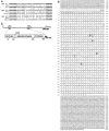
Fig. 1.
Fig. 1.
A, Drosophila melanogaster PCR products with homology to the Kex2 protease family. PCR reactions were conducted on genomic DNA fromDrosophila melanogaster. A translation of the DNA sequence of the three PCR products obtained, DMH #5,DMH #11, and DMH #15, is shown; below each is the Kex2 family member to which it has the greatest amino acid identity: PC2, furin, and Kex2, respectively. Theslashes indicate conceptual splice sites in theDrosophila products at consensus splice sequences and the known splice site in human furin and PC2. The amino acid sequences corresponding to the primers used are printed in bold; the percentage of identical amino acids found in the region between the primers for each Drosophila product and its closest homolog is also indicated. B, The sequence of theDrosophila PC2 cDNA and protein. DMH #5 was used to screen an adult Drosophila head library.A shows the complete nucleotide sequence of the longestamontillado cDNA obtained along with the amino acid sequence of its longest open reading frame. Catalytic triad residues are circled. The aspartate residue unique to PC2s at the position of the subtilisin oxyanion hole asparagine, which is predicted to stabilize the substrate oxyanion during catalysis, isboxed. The residues predicted by molecular modeling to underlie hPC2 specificity for dibasic residues areunderlined and overlined. The predicted signal peptide is underlined; the predicted autoproteolytic cleavage site is indicated by an arrow. These sequence data have been submitted to the DDBJ/EMBLJ/GenBank databases under accession number AF033117. C, A schematic summary indicates the positions of the nucleotides at the beginning and end of the open reading frame, the length of the cDNA and predicted protein, the domain structure of the predicted protein, and the amino acid at which each domain ends.
Fig. 2.
Fig. 2.
Temporal expression pattern ofamontillado RNA during development. The PCR product corresponding to amontillado was hybridized to a developmental Northern blot prepared with 10 μg of total RNA per lane. The lanes correspond to seven different time periods encompassing the first 24 hr of embryogenesis, first and second instar larvae (L1 and L2), early and late third instar larvae (EL and LL), wandering third instar larvae (W3), white prepupae (WP), early, mid, and late pupae (EP, MP, andLP) and female and male adult flies. The Northern blot was subsequently hybridized to RP49 as a loading control, which is known to drop in levels in adult male flies (Al-Atia et al., 1985). The higher expression in adult males as compared with females was not observed in a second independent collection from flies raised in small bottles rather than in larger cages.
Fig. 3.
Fig. 3.
In situ hybridization ofamontillado to embryos. Antisense RNA probes corresponding to half of the amontillado cDNA were hybridized to Drosophila embryos. A, Signal was first observed at stage 15 in a segmentally repeated pattern in the nervous system (arrows). B, During stage 16, signal appears in the brain (hollow arrows) and near the pharynx (filled arrow), in the subesophageal ganglion (∗∗), and at unique positions in the nerve cord (one example shown with ∗). C, During early stage 17 the number of cells expressing in the brain and the intensity of all expressing cells increases. By late stage 17 (shown in Dand E, E being a later time point and different focal plane), staining in the nerve cord can be roughly distinguished at a dorsal, middle, and ventral level (D), and staining begins in the gut (arrow). Sensory structures at the anterior of the embryo also show staining (E, arrows).F, No hybridization was observed at any of these stages with a sense control probe (stage 17 shown). Scale bar, 50 μm.
Fig. 4.
Fig. 4.
amontillado RNA expression in anterior sensory structures during stage 17. A, A lateral view of the anterior of whole-mount stage 17 embryo hybridized to antisense amontillado RNA probe. B, A dorsal view of such a stage 17 embryo. C, A lateral view of a dissected first instar larvae similarly hybridized. Note that the expression in all structures seen in stage 17 is completely absent in L1: in the antennal maxillary complex (thick arrow), the epiphysis (thin arrow), and two other cells lying near the esophagus (hollow arrows). Expression was seen in the attached brain in the first instars examined as a positive control for the staining reactions. Scale bar, 10 μm.
Fig. 5.
Fig. 5.
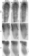
In situ hybridizations ofamontillado to stage 17, first and second instar larval brains. amontillado antisense RNA probes were hybridized to whole-mount stage 17 embryos (A, D,G) and to dissected first (B,E, H) and second (C, F, I) larval instar brains. Dorsal (A–C), midbrain (D–F), and ventral (G–I) focal planes are shown for each of the three different stages from a brain selected for having the greatest number of hybridizing cells of the brains examined from that stage. Note that most of the cells exhibit decreased or no amontillado expression by first instar, and almost all expression is absent by the second larval instar. In D, E, and F,arrows mark some of the positions of cells that undergo such regulation. Arrows in G throughI indicate the two midprotocerebral cells present in each brain hemisphere with consistently strong expression during these three stages. Scale bars, 10 μm.
Fig. 6.
Fig. 6.
In situ hybridizations ofamontillado to stage 17, first and second instar larval ventral nerve cords. amontillado antisense RNA probes were hybridized to whole-mount stage 17 embryos (A–C) and to dissected L1 (D–F) and L2 (G–I) ventral nerve cords. Dorsal (A, D,G), middle (B, E,H), and ventral (C,F, I) focal planes are shown for each of the three stages. Compare A withD and G, and compare Cwith F and I where arrowsindicate some of the cells that show strong amontilladoexpression in stage 17 but severely reduced or abolished expression by L1. In B, E, and H, thesolid arrows indicate the cells in which expression remains strong in L1 but drops in L2. (The third group of cells indicated with a solid arrow in B is in a more dorsal focal plane in the L1 larva, so is seen in Drather than E.) Hollow arrows indicate the cells in which expression has already dropped by L1. The two dark anterior circles in E, F,H, and I are the dorsohaemal appendages and do not represent hybridizing cells. Scale bar, 10 μm.
Fig. 7.
Fig. 7.
Schematic of results of amontillado in situ hybridization to the CNS of stage 17, L1, and L2. The results summarize amontilladoin situhybridization to the CNSs from at least six different larvae in stage 17, L1 and L2. The outlines of the nervous systems are traced from photographs. Scale bar, 25 μm. Each markindicates that expression was observed in a cell at that location in at least one-half of the examined animals. In stage 17, differentcolors and shapes are used to indicate the different focal planes from dorsal to ventral as shown by thekey. Where the identity of cells expressing at later stages could be reasonably assigned based on their similar location, the colored shape was maintained throughout subsequent stages. Lighter colors indicate a lower level of expression. During first and second instar larval stages the location of cells expressing in the brain was quite variable and not always bilaterally symmetric.
Fig. 8.
Fig. 8.
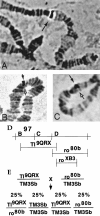
Elimination of the chromosomal region containingamontillado results in a hatching defect. Polytene salivary gland chromosomes obtained from wandering third instar larvae were squashed and hybridized with an amontillado probe. Glands were taken from wild-type Oregon R larvae (A) or from larvae carrying Deletion Df(3R)Tl9QRX(B) or Deletion Df(3R)ro80b (C) over a balancer chromosome. Hybridization was seen on the right arm of the third chromosome in region 97D (filled arrow). Hybridization was absent in the half of the polytene corresponding to the deletion chromosome (hollow arrow), indicating that both deficiencies remove the amontillado gene and their overlap at 97D1–2 contains the amontilladotranscription unit. D, The region from the right arm of the third chromosome from band 97B to 97E is depicted schematically.E, The extent of the genomic region eliminated by Deletion Df(3R) Tl9QRXand Deletion Df(3R) ro80b is indicated by horizontal lines. A cross of two lines that each carry a separate deficiency over a balancer is diagrammed. The expected Mendelian frequencies of the genotypes of the progeny are shown.
Fig. 9.
Fig. 9.
Evidence that amontillado is required for a hatching behavior: larval head swinging.A, The absence of the amontillado region results in a significant reduction in the frequency of head swinging. Stage 17 embryos were videotaped, and the tapes were replayed for analysis. Each head swing is indicated by a vertical line. Hatching is indicated by an arrow. Thehorizontal axis shows increasing time in minutes. The behavioral profiles of three separate individuals from each class are shown, chosen to represent the range of profiles observed. Thetop three graphs are from wild-type Canton S larvae. Themiddle and bottom three graphs are embryos from the cross of Df Tl9QRXand Df ro80b. The middle set corresponds to embryos that hatched, namely DfTl9QRX/TM3Sb or Dfro80b/TM3Sb. The bottom set corresponds to motile nonhatching larvae carrying one copy of each of the two overlapping deletions Df(3R)Tl9QRX/Df(3R)ro80b.B, Expression of wild-type, but not of mutant,amontillado rescues the head-swinging defect. Shown is the distribution of the frequency of swinging behavior in wild-type and mutant embryos with and without amontillado. The number of swings observed within a 3 hr period was counted for 22 heat-shocked Df(3R)Tl9QRX/Df(3R)ro80blarvae, 31 heat-shocked Df(3R)Tl9QRX/Df(3R)ro80blarvae carrying the hs:amon transgene, and 25 heat-shocked Df(3R)Tl9QRX/Df(3R)ro80blarvae carrying the hs:amonH237Atransgene. Twenty wild-type Canton S larvae were analyzed for 3 hr before hatching. The percentage of the population of each strain whose frequency of swinging fell into each of the bins indicated on thex-axis was plotted for each genotype. The mean (±SE) for each of the four different classes, from front to back, was 75 (± 16), 58 (± 13), 147 (± 17), and 275 (± 21).

Similar articles

Cited by

References

    1. Al-Atia GR, Fruscoloni P, Jacobs-Lorena M. Translational regulation of mRNAs for ribosomal proteins during early Drosophila development. Biochemistry. 1985;24:5798–5803. - PubMed
    1. Andrews PC, Brayton KA, Dixon JE. Post-translational proteolytic processing of precursors to regulatory peptides. Experientia [Suppl] 1989;56:192–209. - PubMed
    1. Ashburner M. Drosophila: a laboratory handbook. Cold Spring Harbor Laboratory; Cold Spring Harbor, New York: 1989.
    1. Benjannet S, Rondeau N, Day R, Chretien M, Seidah NG. PC1 and PC2 are proprotein convertases capable of cleaving proopiomelanocortin at distinct pairs of basic residues. Proc Natl Acad Sci USA. 1991;88:3564–3568. - PMC - PubMed
    1. Bernheim SM, Mayeri E. Complex behavior induced by egg-laying hormone in Aplysia. J Comp Physiol [A] 1995;176:131–136. - PubMed

Publication types

Associated data