. 1999 Aug 15;19(16):7007-24.
doi: 10.1523/JNEUROSCI.19-16-07007.1999.

Excess of serotonin (5-HT) alters the segregation of ispilateral and contralateral retinal projections in monoamine oxidase A knock-out mice: possible role of 5-HT uptake in retinal ganglion cells during development

Affiliations

Excess of serotonin (5-HT) alters the segregation of ispilateral and contralateral retinal projections in monoamine oxidase A knock-out mice: possible role of 5-HT uptake in retinal ganglion cells during development

A L Upton et al. J Neurosci. .

Abstract

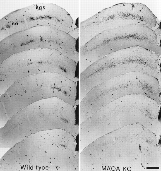
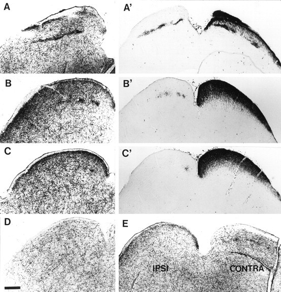
Retinal ganglion cell (RGCs) project to the ipsilateral and contralateral sides of the brain in the dorsal lateral geniculate nucleus (dLGN) and the superior colliculus (SC). Projections from both eyes are initially intermingled until postnatal day 3 (P3) but segregate into eye-specific layers by P8. We report that this segregation does not occur in monoamine oxidase A knock-out mice (MAOA-KO) that have elevated brain levels of serotonin (5-HT) and noradrenaline. The abnormal development of retinal projections can be reversed by inhibiting 5-HT synthesis from P0 to P15. We found that in MAOA-KO mice, 5-HT accumulates in a subpopulation of RGCs and axons during embryonic and early postnatal development. The RGCs do not synthesize 5-HT but reuptake the amine from the extracellular space. In both MAOA-KO and normal mice, high-affinity uptake of 5-HT and serotonin transporter (SERT) immunoreactivity are observed in retinal axons from the optic cup to retinal terminal fields in the SC and dLGN. In the dLGN, transient SERT labeling corresponds predominantly to the ipsilateral retinal projection fields. We show that, in addition to SERT, developing RGCs also transiently express the vesicular monoamine transporter gene VMAT2: thus, retinal axons could store 5-HT in synaptic vesicles and possibly use it as a borrowed neurotransmitter. Finally we show that the 5-HT-1B receptor gene is expressed by RGCs throughout the retina from E15 until adult life. Activation of this receptor is known, from previous studies, to reduce retinotectal activity; thus 5-HT in excess could inhibit activity-dependent segregation mechanisms. A hypothesis is proposed whereby, during normal development, localized SERT expression could confer specific neurotransmission properties on a subset of RGCs and could be important in the fine-tuning of retinal projections.


Figures

Fig. 1.
Fig. 1.
Intraocular injections of HRP in MAOA-KO mice reveal abnormal retinal projections to the dLGN. Sections inA and B are taken from the brain of a 1-month-old wild-type C3H/HeJ mouse and in C andD from a MAOA-KO mouse of the same age. Aand C show the dLGN ipsilateral to the eye injected with HRP. B and D show the dLGN contralateral to the injected eye. A, B, In the wild-type adult, the ipsilateral projection forms dense patches close to the dorsomedial border of the dLGN (A). The contralateral projection fills most of the dLGN, but there is a clear gap that corresponds in size to the patch formed by the ipsilateral projection (B). C,D, In MAOA-KO mice, the ipsilateral projection to the thalamus covers a larger area than in wild-type mice (C); the contralateral projection occupies the entire dLGN and does not contain the characteristic gap (D). Scale bar, 128 μm.
Fig. 2.
Fig. 2.
Quantification of the volume of the ipsilateral projection to the dLGN in wild-type and MAOA-KO mice. To estimate the volume of the dLGN receiving projections from the ipsilateral eye, the area of the dLGN covered by HRP reaction product in each section after intraocular injection of HRP, was measured using an image analysis program. The volume occupied by the terminals from the injected eye was calculated by multiplying the area measured by the thickness of each section (40 μm) from serial coronal sections through the dLGN. Totals are shown in cubic millimeters for wild-type C3H/HeJ mice at P3 (n = 4; mean ± SEM = 0.0102 ± 0.0013), P8 (n = 3; mean ± SEM = 0.0085 ± 0.0018), and P30 (n = 7; mean ± SEM = 0.0151 ± 0.0007) (white bars); for MAOA-KO mice at P3 (n = 4; mean ± SEM = 0.0094 ± 0.0013), P8 (n = 4; mean ± SEM = 0.0192 ± 0.0024), and P30 (n = 4; mean ± SEM = 0.0235 ± 0.0009) (black bars); and for adult MAOA-KO mice treated with PCPA during the first 15 postnatal days (n = 2; mean ± SEM = 0.01527 ± 0.0008) (striped bar). Values for MAOA-KO mice that differ significantly from wild-type are indicated by an asterisk above the bar (p ≤ 0.01, using Student’s unpairedt test). At P3, the ipsilateral projections of wild-type and MAOA-KO mice occupy the same volume of the dLGN, whereas at P8 and P30, the ipsilateral projection in MAOA-KO mice is significantly larger than in wild-type animals. The ipsilateral projections of MAOA-KO mice treated with PCPA occupy the same volume as those of wild-type mice. Error bars indicate the SEM for each value.
Fig. 3.
Fig. 3.
Abnormal retinal projections to the superior colliculus in MAOA-KO mice. One-month-old C3H/HeJ and MAOA-KO mice received intraocular HRP injections and were sacrificed 24 hr later. Rostrocaudal series of one in four, 40 μm coronal sections through the SC are illustrated. In the SC of wild-type animals, the ipsilateral projection forms discernible patches in the stratum opticum (so) and lower stratum griseum superficiale (sgs), and tends to cluster in a medial patch caudally; only scattered axons are visible in the upper SGS. In the SC of MAOA-KO mice, the ipsilateral projection appears to be more diffuse: it does not form patches in the SO, and it has a wider extension dorsally in the SGS, and caudally where it continues to be distributed mediolaterally even at caudal SC levels. Scale bar, 418 μm.
Fig. 4.
Fig. 4.
Development of retinal projections to the dLGN in wild-type and MAOA-KO mice. Intraocular injections of HRP into wild-type C3H/HeJ (+/+) animals aged P3 (A,B) and P8 (E, F) and MAOA-KO animals aged P3 (C, D) and P8 (G, H). A,C, E, and G show the dLGN ipsilateral to the eye injected with HRP. B,D, F, and H show the dLGN contralateral to the eye injected with HRP. A,B, In wild-type mice at P3, retinal projections fill a large proportion of the ipsilateral dLGN (A) and the entire contralateral dLGN (B).C, D, Ipsilateral and contralateral retinal projections illustrated here in MAOA-KO mice at P3 are identical to those of wild-type mice. E,F, In wild-type mice at P8, ipsilateral projections are confined to the dorsomedial part of the dLGN (E), and the contralateral projection has withdrawn from this region (F): segregation of the two inputs appears complete in normal animals. G, H, In MAOA-KO mice at P8, fibers to the ipsilateral (G) and contralateral (H) thalamus still innervate overlapping territories: segregation has failed to occur. Scale bar, 110 μm.
Fig. 5.
Fig. 5.
5-HT immunolabeling in RGCs and their axons during development. 5-HT immunolabeling of the developing visual pathway is readily visible in MAOA-KO mice (A–F), and faint 5-HT immunoreactivity is also detectable in retinal-like fibers in wild-type C3H/HeJ mice (G–I). 5-HT immunolabeling (DAB–nickel staining) appears dark purple. Some sections were counterstained with methyl green (blue and green) to show the histological structures. A–C, In the retina, where the pigment epithelium appears black, 5-HT immunostaining is visible in the ganglion cell bodies in the peripheral ventral retina, shown here at E17 (A, B) and P3 (C). Stained axons can be followed at the surface of the retinal ganglion cell layer (B, C) and in the optic nerve (on) (A, B).D, E, As shown in an E17 embryo, intense 5-HT immunolabeling can be followed along the visual pathway: in the optic tract (ot), in the superficial fibers coursing above the dLGN (D) and in the SC (E). F, A similar pattern is visible in MAOA-KO mice postnatally, until P15, as illustrated here at P8. There is dense 5-HT immunostaining of the entire superficial layers of the SC that ends abruptly at the junction between the superior and inferior colliculi (IC), similarly to the retinal inputs.G–I, In wild-type mice, light 5-HT immunolabeling could occasionally be detected in the visual pathway. G, A dense patch of 5-HT labeling (arrow) is visible in the dLGN of a P6 mouse. The localization of this patch resembles the location of the ipsilateral retinal projection shown in Figure 4G. H,A higher magnification of the same section shows that this patch contains a dense cluster of weakly labeled fine fibers (arrow), contrasting with the well-delineated but sparsely distributed 5-HT-positive fibers, that correspond to fibers originating in the raphe. I, 5-HT-labeled axons tipped with growth cones can be seen advancing in the optic tract in an E17 mouse. Scale bar (in F): A, 750 μm;B, C, 190 μm; D, 300 μm; E, 260 μm; F, 380 μm; G, 600 μm; H, 55 μm; I, 25 μm.
Fig. 6.
Fig. 6.
5-HT* uptake in retinal fibers innervating the superior colliculus of wild-type mice. A–E,High-affinity uptake of tritiated 5-HT in 70-μm-thick vibratome slices of C3H/HeJ mice aged P10. Labeling was revealed by autoradiography after emulsion coating. A′–C′, HRP labeling after injection into the left eye in a C3H/HeJ mouse aged P8.A–C illustrate three coronal sections from rostral (A) to caudal (C) through the superior colliculus. Only one colliculus is shown, and the midline is to the right. High-affinity 5-HT uptake is seen in two different locations: (1) in varicose fibers, distributed all over the brainstem, that correspond to fibers from the raphe; and (2) as dense accumulation of terminals in the superficial SGS in which they form a continuous band, and in the SO and lower SGS in which they form discrete patches caudally (B, C) and a continuous line rostrally (A).A′–C′, The distribution of retinal projections, labeled by intraocular injection of HRP, at three equivalent coronal levels to those in A–C is shown: ipsilateral projections can be observed in the left SC, and contralateral projections in the right SC.D, In a P10 mouse binocularly enucleated on P8, the areas of dense 5-HT uptake have completely disappeared in the SO and SGS of the SC (only one side is illustrated, the level corresponds approximately to that shown in B). E, In a P10 mouse in which the left eye was removed on P8, the area of dense 5-HT uptake is reduced contralaterally to the lesion in the SGS and ipsilaterally to the enucleation in the SO. Scale bar, 340 μm.
Fig. 7.
Fig. 7.
SERT immunolabeling on RGC axons during development in wild-type mice. SERT immunolabeling is visible on RGC axons but not cell bodies in the developing visual pathway.A–D, Coronal cryostat sections (20 μm) from an E17 normal mouse. A, SERT-positive fibers are seen leaving the retina in the ventral part of the optic nerve. B–D,SERT immunolabeling can be followed in the optic nerve (on) (B), the chiasm (C), and the optic tract (ot) (D). Note that labeling is restricted to peripheral parts of the optic nerve. E–J, Free-floating sections from mice aged P8–P10. E, SERT immunolabeling is detected in a limited region of the dLGN (indicated by anarrow) as well as in thalamic axons in the reticularis (rt). F, G, After injections of HRP into one eye at P8, alternate brain sections were treated either to reveal the HRP (G) or immunolabeled for SERT (F). Exact superposition of adjacent sections was not possible because the two procedures resulted in different degrees of shrinkage of the tissue. However, alignment of consecutive sections indicated that the region of SERT immunolabeling in the dLGN corresponds to the area of the gap in contralateral projections (G) that receives projections from the ipsilateral eye. H,I, After monocular enucleation at P8, SERT-labeled fibers at P10 have totally disappeared from the dLGN ipsilateral to the eye that has been removed (I) but are still present ipsilateral to the remaining eye (i.e., contralateral to the enucleation) (H). J, In the SC at P9, SERT expression is concentrated in the SO in patches reminiscent of the ipsilateral retinal innervation and is not detectable in more superficial SGS. K, L, In higher magnification micrographs of SERT labeling at P0 in the optic tract (K) and at the pretectal–SC junction (L) raphe fibers appear densely immunoreactive and varicose, whereas the presumptive SERT-positive optic fibers are more linear, more lightly labeled, and are grouped in bundles. Scale bar: A, 331 μm; B–D, 230 μm;E, J, 400 μm; F,G, 150 μm; H, I, 300 μm; K, L, 35 μm.
Fig. 8.
Fig. 8.
In situ hybridization of SERT, 5-HT1B receptor, and VMAT2. Antisense cRNA35S-labeled probes to SERT (B,F, J), the 5-HT1Breceptor (C, G, K) and VMAT2 (D, H, L) were hybridized to 15-μm-thick coronal sections through the retina at E15 (AD), P1 (E–H), and P6 (I–L). The retinal pigmented epithelium (pe) was nonspecifically labeled by sense and antisense probes. A, E, andI are bright-field photomicrographs of Nissl-stained sections. A, At E15, two layers can be distinguished in the retina: the undifferentiated neuroepithelial cells and the postmitotic retinal ganglion cell layer (gc). The latter, outlined by a dashed line, first appears close to the optic nerve head (O) and subsequently at the periphery. E, At P1, the ganglion cell layer is clearly separated from the others by the inner plexiform layer.I, By P6, differentiation of the different retinal cell types is complete, and all layers of the retina are distinguishable.B–D, At E15, a small area at the periphery of the retinal ganglion cell (RGC) layer is positive for SERT (B), and a hybridization signal is observed in the entire RGC layer with probes for 5-HT1B(C) and VMAT2 (D).F–H, At P1, a restricted territory at the periphery of the RGC layer expresses SERT (F), whereas the entire RGC layer is still labeled with 5-HT1B(G) and VMAT2 (H).J–L, At P6, SERT mRNA labeling has become confined to small patches in the ventral part of the peripheral retina (J). 5-HT1B expression still extends throughout the RGC layer (K), whereasVMAT2 labeling (L) starts to decrease compared with younger ages. Scale bar: A–D, I–L, 150 μm; E–H, 300 μm.
Fig. 9.
Fig. 9.
SERT expression in relationship to ipsilateral retinal projections. A, B, Coronal sections taken through the retina of a P4 mouse pup injected in the ipsilateral thalamus on P1 with fluorogold. A, ISH using a probe for SERT mRNA. An arrow indicates the furthest extent of the ISH signal in the RGC layer. B, Same section photographed under UV fluorescence to reveal fluorogold retrogradely transported to the retina (magnification, 44×). Thearrow indicates the edge of the ipsilaterally projecting region. Note that SERT hybridization signal in the ventral retina falls within the region containing ipsilaterally projecting cells.C, A reconstruction of the retina was made from a series of 15 μm horizontal sections, 60 μm apart, taken through one E17 C3H/HeJ retina and processed for ISH. The circumference of each retinal section is represented as a straight gray line with the region of SERT expression in RGCs represented by a thicker green bar. In sections through the lens, the end of the ganglion cell layer was taken as the end of the line. However, the most dorsal and the three most ventral sections that contained a continuous ganglion cell layer were divided at the point closest to the surface of the skin for the purpose of this reconstruction. The position of the optic nerve is indicated by a filled circle. The labeled territory forms a peripheral crescent that only excludes the dorsal peripheral retina. D, Summary of the anatomical organization of the crossed and uncrossed retinal pathways in rodents as previously described in the literature (Dräger and Olson, 1980; Métin et al., 1983; Reese and Cowey, 1983; Godement et al., 1984). The ipsilateral projections, shown in red, originate in the ventrotemporal crescent of the retina, covering 20% of the retinal surface. Ipsilateral terminals are essentially clustered in the medial dLGN and in the SO and lower SGS of the SC. Contralateral projections to the SC originate from the entire retina (blue). Contralateral projections overlap with ipsilateral projections in the rostral SC (designated by an intermediate violet color) but not in the dLGN. E, Summary diagram of the transient localization of SERT observed in the present study. This includes data obtained by ISH in the retina, and SERT immunocytochemistry and high-affinity 5-HT uptake in the terminal fields. The area of SERT expression, shown in green, covers the periphery of the retina, excluding only the dorsalmost region. In the dLGN, SERT is present in the ipsilateral terminal field. In the SC, high-affinity uptake is present in the ipsilateral zones of the SO and lower SGS, but also in contralateral fibers innervating the top part of the SGS.

Similar articles

Cited by

References

    1. Bennett-Clarke CA, Leslie MJ, Chiaia NL, Rhoades RW. Serotonin 1B receptors in the developing somatosensory and visual cortices are located on thalamocortical axons. Proc Natl Acad Sci USA. 1993;90:153–157. - PMC - PubMed
    1. Bennett-Clarke CA, Leslie MJ, Lane RD, Rhoades RW. Effect of serotonin depletion on vibrissa-related patterns of thalamic afferents in the rat’s somatosensory cortex. J Neurosci. 1994;14:7594–7607. - PMC - PubMed
    1. Bhide PG, Frost DO. Stages of growth of hamster retinofugal axons: implications for developing axonal pathways with multiple targets. J Neurosci. 1991;11:485–504. - PMC - PubMed
    1. Blakely RD, Berson HE, Fremeau RT, Jr, Caron MG, Peek MM, Prince HK, Bradley CC. Cloning and expression of a functional serotonin transporter from rat brain. Nature. 1991;354:66–70. - PubMed
    1. Blue ME, Erzurumlu RS, Jhaveri S. A comparison of pattern formation by thalamocortical and serotonergic afferents in the rat barrel-field cortex. Cereb Cortex. 1991;1:380–389. - PubMed

Publication types

MeSH terms

LinkOut - more resources