

Changes in neuropeptide Y receptors and pro-opiomelanocortin in the anorexia (anx/anx) mouse hypothalamus
- PMID: 10436066
- PMCID: PMC6782872
- DOI: 10.1523/JNEUROSCI.19-16-07130.1999
Changes in neuropeptide Y receptors and pro-opiomelanocortin in the anorexia (anx/anx) mouse hypothalamus
Abstract
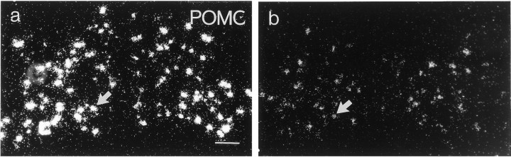
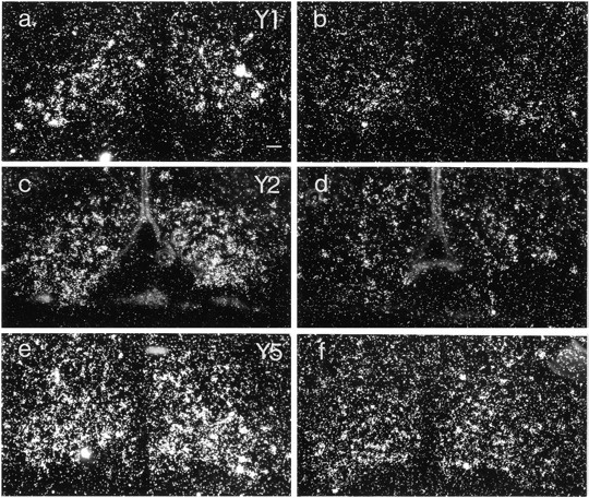
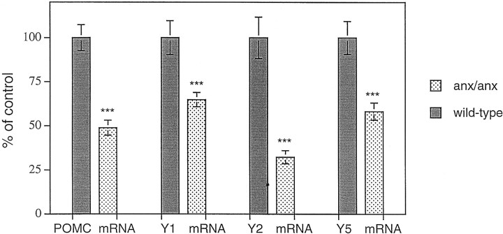
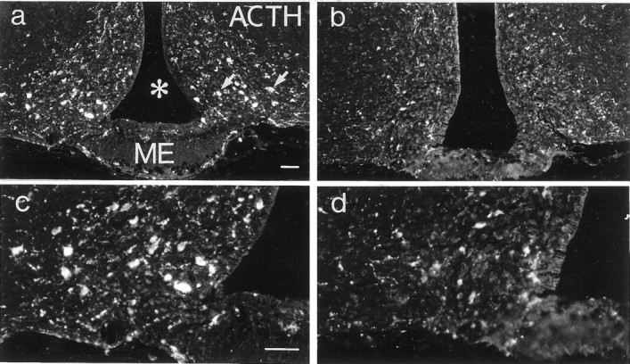
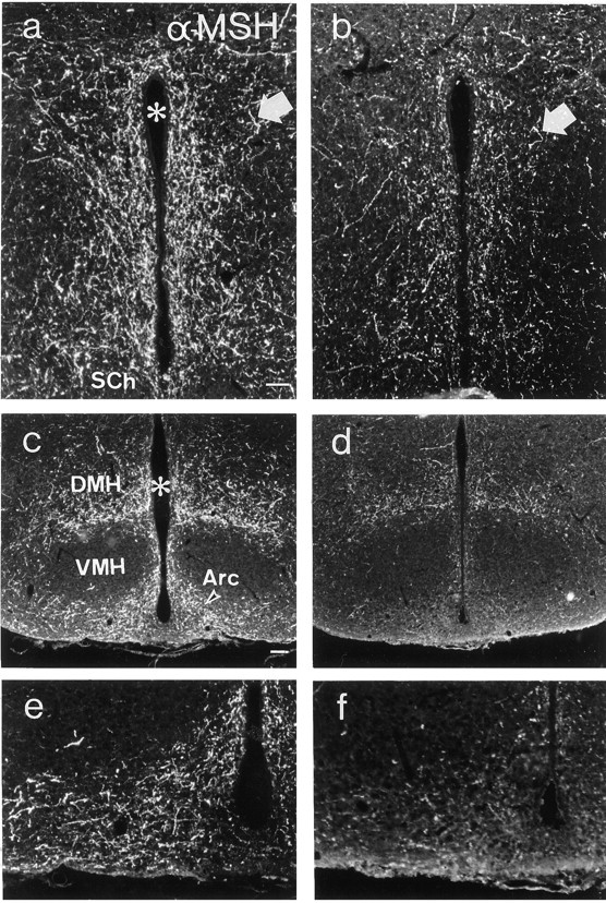
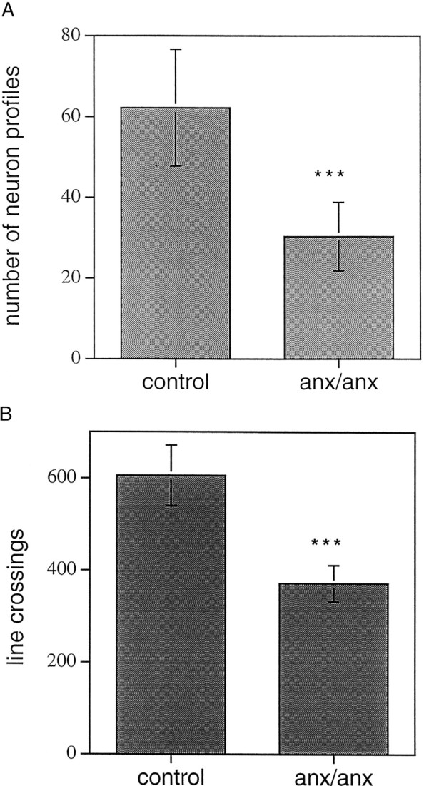
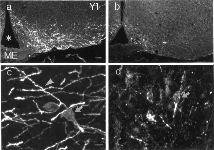
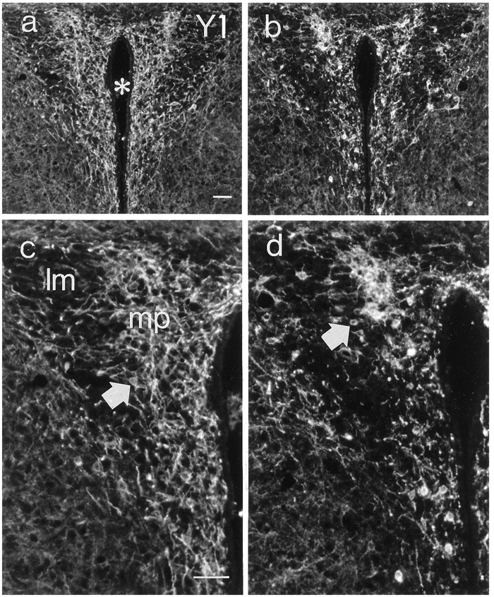
The pro-opiomelanocortinergic (POMCergic) system originating in the hypothalamic arcuate nucleus extends projections widely over the brain and has been shown to be intricately linked and parallel to the arcuate neuropeptide Y (NPY) system. Both NPY and POMC-derived peptides (melanocortins) have been strongly implicated in the control of feeding behavior, with the former exerting orexigenic effects and the latter having anorexigenic properties. Mice homozygous for the lethal anorexia (anx) mutation are hypophagic, emaciated, and exhibit anomalous processing of NPY exclusively in the arcuate nucleus, providing an interesting model to study NPY-POMC interactions. In the present study, several morphological markers were used to investigate the histochemistry and morphology of the POMC system in anx/anx mice. In situ hybridization demonstrated decreased numbers of POMC mRNA-expressing neurons in the anx/anx arcuate nucleus. In parallel, mRNA levels for both the NPY Y1 and Y5 receptors, which are expressed in POMC neurons, were decreased. Also, expression of the NPY Y2 autoreceptor was attenuated. Immunohistochemistry using antibodies against adrenocorticotropic hormone to demonstrate POMC cell bodies, against alpha-melanocyte-stimulating hormone to demonstrate axonal projections and against the NPY Y1 receptor to demonstrate dendritic arborizations, showed strikingly decreased immunoreactivities for all these markers. The present data suggest that degeneration of the arcuate POMC system is a feature characteristic of the anx/anx mouse. The possible relationship to the NPYergic phenotype of this animal is discussed.
Figures
Similar articles
-
Alterations of arcuate nucleus neuropeptidergic development in contactin-deficient mice: comparison with anorexia and food-deprived mice.Eur J Neurosci. 2005 Dec;22(12):3217-28. doi: 10.1111/j.1460-9568.2005.04513.x. Eur J Neurosci. 2005. PMID: 16367788
-
Subtypes Y1 and Y2 of the neuropeptide Y receptor are respectively expressed in pro-opiomelanocortin- and neuropeptide-Y-containing neurons of the rat hypothalamic arcuate nucleus.Neuroendocrinology. 1997 Dec;66(6):393-408. doi: 10.1159/000127265. Neuroendocrinology. 1997. PMID: 9430445
-
Evidence of hypothalamic degeneration in the anorectic anx/anx mouse.Glia. 2011 Jan;59(1):45-57. doi: 10.1002/glia.21075. Epub 2010 Oct 21. Glia. 2011. PMID: 20967882
-
Neuropeptide Y: some viewpoints on a multifaceted peptide in the normal and diseased nervous system.Brain Res Brain Res Rev. 1998 May;26(2-3):154-66. doi: 10.1016/s0165-0173(97)00052-0. Brain Res Brain Res Rev. 1998. PMID: 9651513 Review.
-
Molecular analysis of central feeding regulation by neuropeptide Y (NPY) neurons with NPY receptor small interfering RNAs (siRNAs).Neurochem Int. 2012 Nov;61(6):936-41. doi: 10.1016/j.neuint.2012.02.029. Epub 2012 Mar 5. Neurochem Int. 2012. PMID: 22414532 Review.
Cited by
-
Hypothalamic integration of immune function and metabolism.Prog Brain Res. 2006;153:367-405. doi: 10.1016/S0079-6123(06)53022-5. Prog Brain Res. 2006. PMID: 16876587 Free PMC article. Review.
-
The anx/anx Mouse - A Valuable Resource in Anorexia Nervosa Research.Front Neurosci. 2019 Feb 5;13:59. doi: 10.3389/fnins.2019.00059. eCollection 2019. Front Neurosci. 2019. PMID: 30804742 Free PMC article. Review.
-
Hypothalamic mitochondrial dysfunction associated with anorexia in the anx/anx mouse.Proc Natl Acad Sci U S A. 2011 Nov 1;108(44):18108-13. doi: 10.1073/pnas.1114863108. Epub 2011 Oct 24. Proc Natl Acad Sci U S A. 2011. PMID: 22025706 Free PMC article.
-
The use of animal models to decipher physiological and neurobiological alterations of anorexia nervosa patients.Front Endocrinol (Lausanne). 2015 May 19;6:68. doi: 10.3389/fendo.2015.00068. eCollection 2015. Front Endocrinol (Lausanne). 2015. PMID: 26042085 Free PMC article. Review.
-
AgRP neurons trigger long-term potentiation and facilitate food seeking.Transl Psychiatry. 2021 Jan 5;11(1):11. doi: 10.1038/s41398-020-01161-1. Transl Psychiatry. 2021. PMID: 33414382 Free PMC article.
References
-
- Bai FL, Yamano M, Shiotani Y, Emson PC, Smith AD, Powell JF, Tohyama M. An arcuato-paraventricular and -dorsomedial hypothalamic neuropeptide Y-containing system which lacks noradrenaline in the rat. Brain Res. 1985;331:172–175. - PubMed
-
- Barnea A, Cho G, Porter JC. Intracellular processing of alpha-MSH and ACTH in hypothalamic neurons: a preliminary study. Soc Neurosci Abstr. 1979;5:523.
-
- Brady LS, Smith MA, Gold PW, Herkenham M. Altered expression of hypothalamic neuropeptide mRNAs in food-restricted and food-deprived rats. Neuroendocrinology. 1990;52:441–447. - PubMed
-
- Broberger C, Johansen J, Schalling M, Hökfelt T. Hypothalamic neurohistochemistry of the murine anorexia (anx/anx) mutation: Altered processing of neuropeptide Y (NPY) in the arcuate nucleus. J Comp Neurol. 1997a;387:124–135. - PubMed
-
- Broberger C, Landry M, Wong H, Walsh J, Hökfelt T. Subtypes Y1 and Y2 of the neuropeptide Y receptor are respectively expressed in proopiomelanocortin and neuropeptide Y-containing neurons of the rat hypothalamic arcuate nucleus. Neuroendocrinology. 1997b;66:393–408. - PubMed
Publication types
MeSH terms
Substances
Grants and funding
LinkOut - more resources
Full Text Sources
Molecular Biology Databases
Research Materials
Miscellaneous