

Review
doi: 10.1128/JB.181.16.4725-4733.1999.
Structures of gram-negative cell walls and their derived membrane vesicles
Affiliations
- PMID: 10438737
- PMCID: PMC93954
- DOI: 10.1128/JB.181.16.4725-4733.1999
Item in Clipboard
Review
Structures of gram-negative cell walls and their derived membrane vesicles
J Bacteriol.
1999 Aug.
No abstract available
Figures
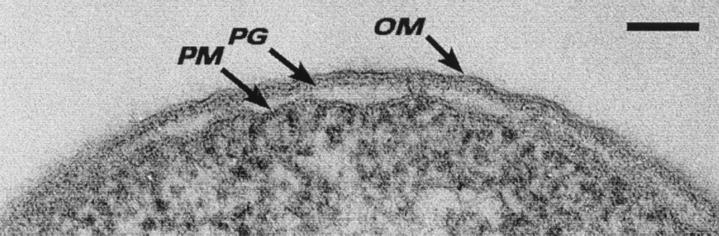
Thin section of the cell envelope of E. coli K-12 after conventional embedding. The periplasmic space is empty of substance, and the peptidoglycan layer (PG), outer membrane (OM), and plasma membrane (PM) can be seen. Bar = 100 nm.
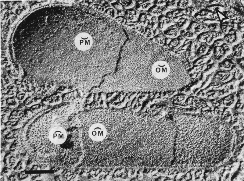
Freeze-etching of two E. coli K-12 cells in which the fracture planes have travelled through the cell envelope. The upper cell shows concave fractures through the outer membrane (OM) and plasma membrane (PM), whereas the lower cell shows convex fractures of the same membranes. The particles (or holes) in these membrane fractures correspond to intramembranous protein complexes. The arrowhead in the upper right-hand corner of the image points out the shadow direction. Bar = 500 nm. (Reprinted from reference .)
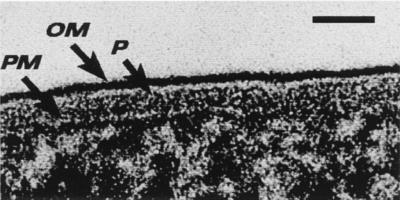
Freeze-substitution image of the E. coli K-12 cell envelope for direct comparison with Fig. 1. The periplasmic space is filled with periplasm (P) (the so-called periplasmic gel), and the peptidoglycan layer is not visible. The outer face of the outer membrane (OM) is densely stained, because the LPS retains its asymmetric location in this region of the bilayer and is more highly charged than the phospholipid on the inner face of the OM. The periplasm is bounded by the OM and the plasma membrane (PM). Bar = 25 nm. (Reprinted from reference .)
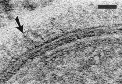
Freeze-substitution image of the P. aeruginosa PAO1 cell envelope showing the long O-side chains of the B-band LPS extending ∼40 nm from the face of the outer membrane (arrow). Bar = 35 nm. (Reprinted from reference .)
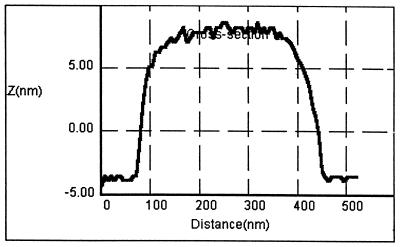
AFM tracing over a single hot-phenol-extracted A- and B-band LPS micelle from P. aeruginosa PAO1 obtained under water (pH 7.0) after the micelle had attached to a silicon nitride surface. The 12-nm height (Z on the y axis) of the vesicle represents both the lower and upper faces of the LPS bilayer and is consistent with only the lipid A plus the core oligosaccharide being detected on the 300-nm-diameter micelle. Yet, sodium dodecyl sulfate-polyacrylamide gel electrophoresis of the preparation showed that the O-side chains of both LPSs are present, and freeze-substitution images of intact outer membrane surfaces (Fig. 4) revealed that the B-band O-side chain extends ∼40 nm. We believe the O-side chains are so rapidly moving under the aqueous conditions of our AFM experiment that the AFM tip cannot detect them.
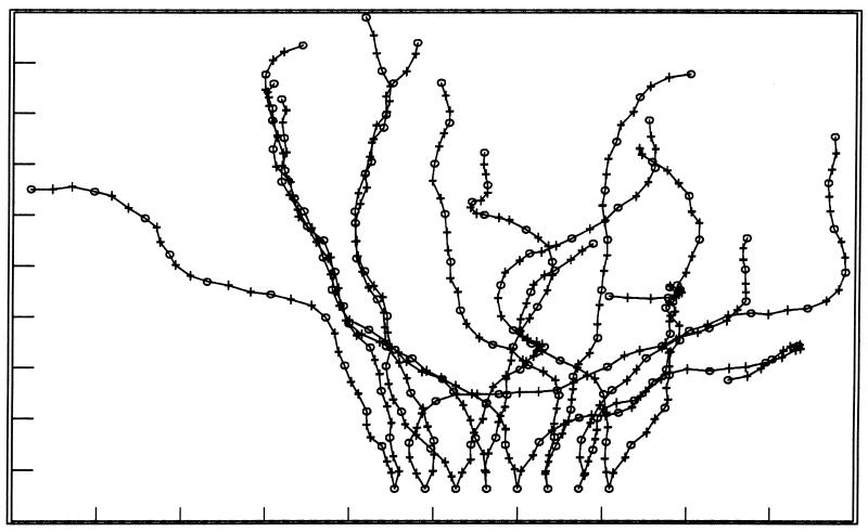
Schematic diagram showing the different alignments (i.e., movement) of the B-band O-side chains of P. aeruginosa as we perceive them from the AFM experiments shown in Fig. 5 (53). The model assumes that all ionizable groups are charged. The core oligosaccharide and lipid A moieties are not shown but would be at the bottom of the diagram.
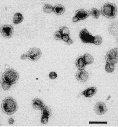
Negative-stained n-MVs which have been isolated and purified from P. aeruginosa as previously described (32). Bar = 250 nm.
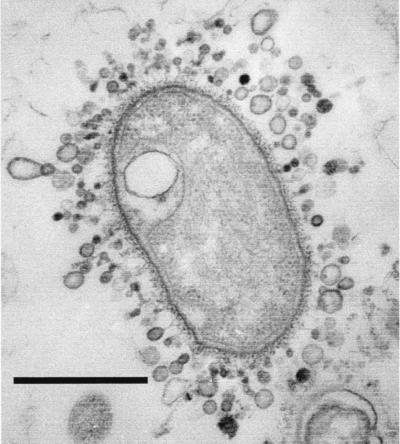
Thin section of an unidentified gram-negative bacterium found in a freshwater biofilm in a river near laboratory. This bacterium possesses a microcapsule and is liberating a prodigious amount of MVs. Bar = 1 μm.
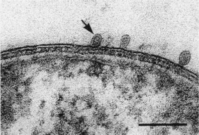
Thin section of P. aeruginosa PAO1 showing the development of n-MVs before they are liberated from the cell. The arrow points to one vesicle in which the membrane bilayer and the periplasm within its lumen (i.e., electron-dense area inside the vesicle) can be seen. Bar = 250 nm.
References
-
- Amako K, Murata K, Umeda A. Structure of the envelope of Escherichia coli observed by the rapid freezing and substitution fixation method. Microbiol Immunol. 1983;27:95–99. - PubMed
-
- Bayer M E. Zones of membrane adhesion in the cryofixed envelope of Escherichia coli. J Struct Biol. 1991;197:268–280. - PubMed
-
- Beveridge T J. Ultrastructure, chemistry, and function of the bacterial cell wall. Int Rev Cytol. 1981;72:229–317. - PubMed
Publication types
MeSH terms
Substances
LinkOut - more resources
Full Text Sources
Other Literature Sources
