. 1999 Aug;181(16):4825-33.
doi: 10.1128/JB.181.16.4825-4833.1999.

Transformations in flagellar structure of Rhodobacter sphaeroides and possible relationship to changes in swimming speed

Affiliations

Transformations in flagellar structure of Rhodobacter sphaeroides and possible relationship to changes in swimming speed

J P Armitage et al. J Bacteriol. 1999 Aug.

Abstract

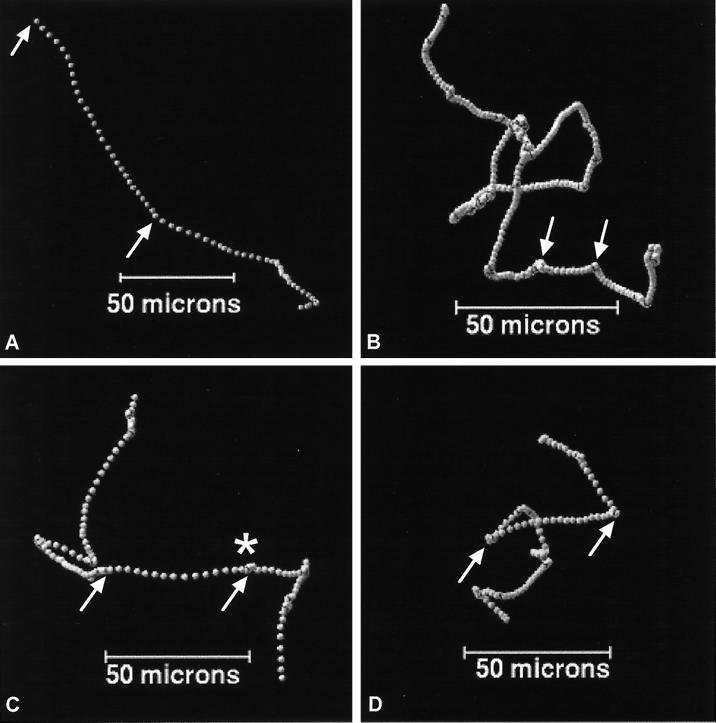
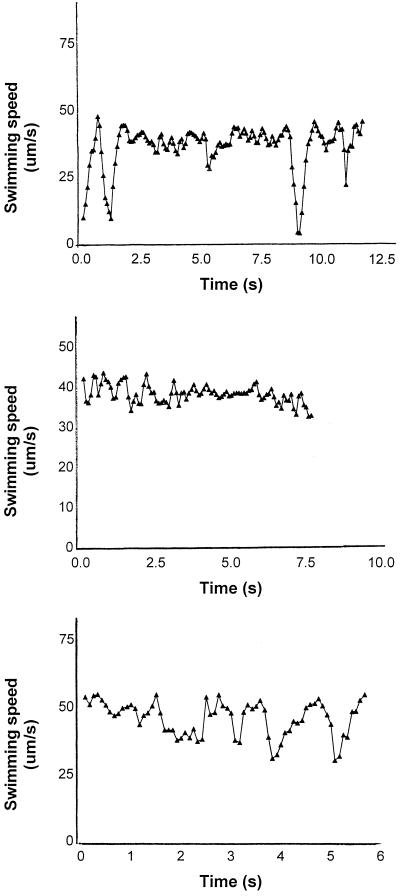
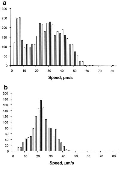
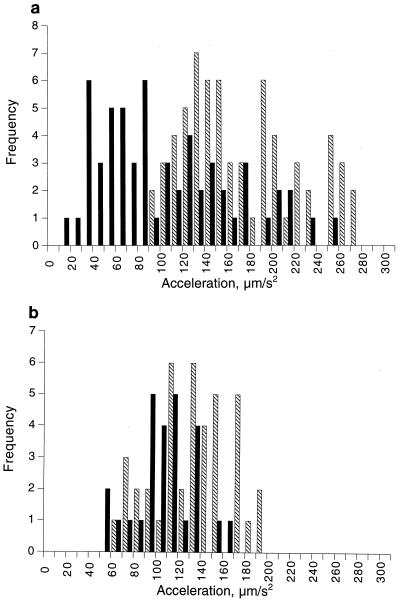
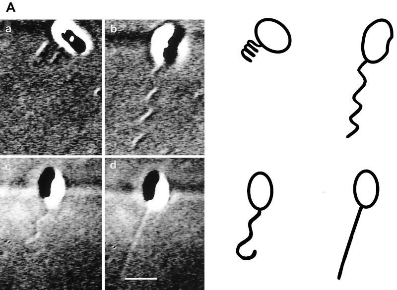
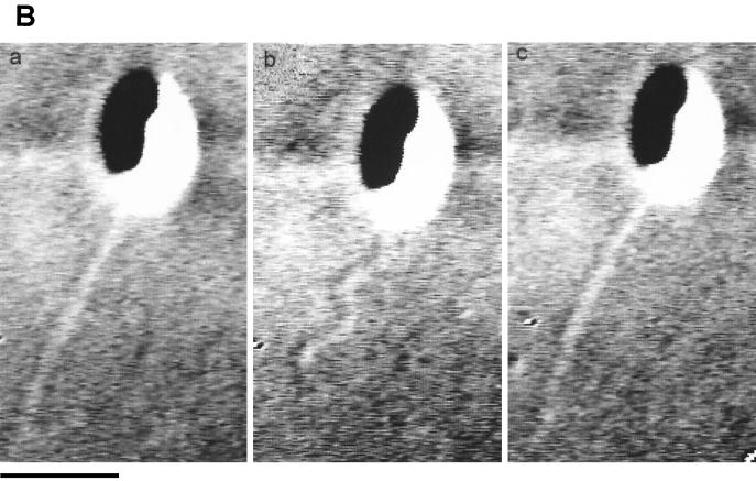
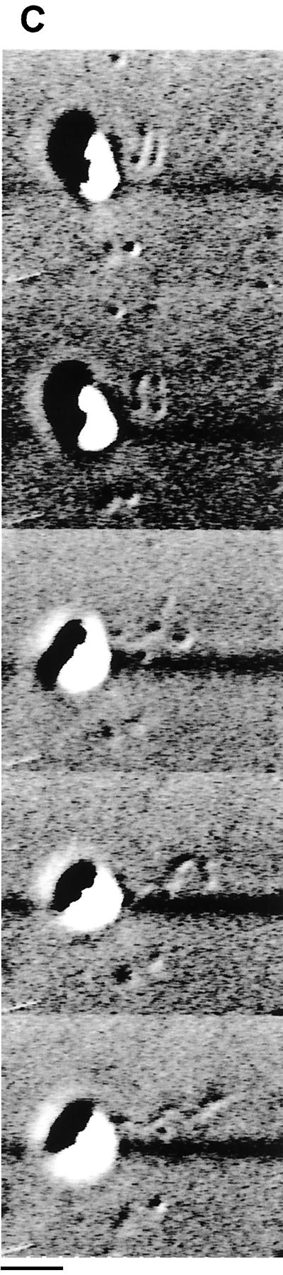
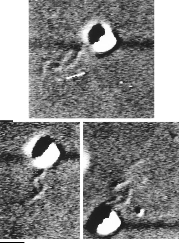
Rhodobacter sphaeroides is a photosynthetic bacterium which swims by rotating a single flagellum in one direction, periodically stopping, and reorienting during these stops. Free-swimming R. sphaeroides was examined by both differential interference contrast (DIC) microscopy, which allows the flagella of swimming cells to be seen in vivo, and tracking microscopy, which tracks swimming patterns in three dimensions. DIC microscopy showed that when rotation stopped, the helical flagellum relaxed into a high-amplitude, short-wavelength coiled form, confirming previous observations. However, DIC microscopy also revealed that the coiled filament could rotate slowly, reorienting the cell before a transition back to the functional helix. The time taken to reform a functional helix depended on the rate of rotation of the helix and the length of the filament. In addition to these coiled and helical forms, a third conformation was observed: a rapidly rotating, apparently straight form. This form took shape from the cell body out and was seen to form directly from flagella that were initially in either the coiled or the helical conformation. This form was always significantly longer than the coiled or helical form from which it was derived. The resolution of DIC microscopy made it impossible to identify whether this form was genuinely in a straight conformation or was a low-amplitude, long-wavelength helix. Examination of the three-dimensional swimming pattern showed that R. sphaeroides changed speed while swimming, sometimes doubling the swimming speed between stops. The rate of acceleration out of stops was also variable. The transformations in waveform are assumed to be torsionally driven and may be related to the changes in speed measured in free-swimming cells. The roles of and mechanisms that may be involved in the transformations of filament conformations and changes in swimming speed are discussed.


Figures

FIG. 1
FIG. 1
Three-dimensional swimming patterns of three individual R. sphaeroides cells (A, B, and C) and one E. coli cell (D). Between the arrows, the plots are oriented so that the cell is swimming parallel to the plane of view. Each spot represents 1/12 s. The closer together the spots, the slower the cell speed. The cells in panels A and D were swimming top to bottom, while those in panels B and C were swimming bottom to top. The asterisk marks a stop followed by acceleration out of that stop.
FIG. 2
FIG. 2
Change in swimming speed of three individual R. sphaeroides cells tracked in three dimensions. When the speed falls below 10 μm s−1, the cells have probably stopped, and the measured speed probably represents Brownian motion, although in some cases there may be slow movement. The time scales are different, as the cells were tracked for different periods of time before being lost from tracking.
FIG. 3
FIG. 3
Distribution of swimming speed during periods of smooth swimming between stops, calculated from 32 tracked R. sphaeroides cells (a) and 11 tracked E. coli cells (b). Below 10 μm s−1, R. sphaeroides cells were considered stopped. The number of the y axis refers to the number of periods of smooth swimming, and the speed on the x axis is the average speed during a period of smooth swimming.
FIG. 4
FIG. 4
Distribution of acceleration rates of 32 R. sphaeroides cells tracked from stop to swim (▧) and speed changes while free swimming (■) (a) and 11 tracked E. coli cells (b) (symbols are as in panel a). The frequency with which the individual acceleration rates were measured (as described in Materials and Methods) is shown on the y axis. Data show 95% confidence limits. The averages (mean ± standard deviation) of the behaviors measured over the entire histogram were as follows: ▧, 178 ± 18 and 122 ± 10 μm s−2 for R. sphaeroides and E. coli, respectively; ■, 95 ± 15 and 99 ± 10 μm s−2 for R. sphaeroides and E. coli, respectively.
FIG. 5
FIG. 5
DIC images of the flagellar filament of R. sphaeroides. (A) Various flagellar conformations: a coiled form (a), a functional helix (b), a helix relaxing into a coil (c), and an apparently straight conformation (d). The cartoon shows the likely cell size and the flagellar shape. (B) Filament changing between straight and helical conformations. (C) Sequential formation of a functional helix from a coiled filament. The two images at the top show the coiled form rotating. Bars, 1 μm.
FIG. 5
FIG. 5
DIC images of the flagellar filament of R. sphaeroides. (A) Various flagellar conformations: a coiled form (a), a functional helix (b), a helix relaxing into a coil (c), and an apparently straight conformation (d). The cartoon shows the likely cell size and the flagellar shape. (B) Filament changing between straight and helical conformations. (C) Sequential formation of a functional helix from a coiled filament. The two images at the top show the coiled form rotating. Bars, 1 μm.
FIG. 6
FIG. 6
DIC images of a dividing cell with two independently rotating flagellar filaments. The two flagella rotated independently, even when close together. Bar, 1 μm.

References

    1. Adler J, Dahl L. A method for measuring the motility of bacteria and comparing random and non-random motility. J Gen Microbiol. 1967;46:161–173. - PubMed
    1. Amsler C D, Matsumura P. Chemotactic signal transduction in Escherichia coli and Salmonella typhimurium. In: Hoch J A, Silhavy T J, editors. Two-component signal transduction. Washington, D.C: American Society for Microbiology; 1995. pp. 89–103.
    1. Armitage J P. Bacterial motility and chemotaxis. Sci Prog. 1992;76:451–477. - PubMed
    1. Armitage J P, Macnab R M. Unidirectional intermittent rotation of the flagellum of Rhodobacter sphaeroides. J Bacteriol. 1987;169:514–518. - PMC - PubMed
    1. Armitage J P, Schmitt R. Bacterial chemotaxis: variations on a theme: Rhodobacter sphaeroides and Sinorhizobium meliloti. Microbiology. 1997;143:3671–3682. - PubMed

Publication types

MeSH terms

LinkOut - more resources