. 1999 Aug;181(16):4937-48.
doi: 10.1128/JB.181.16.4937-4948.1999.

Excision of IS492 requires flanking target sequences and results in circle formation in Pseudoalteromonas atlantica

Affiliations

Excision of IS492 requires flanking target sequences and results in circle formation in Pseudoalteromonas atlantica

D Perkins-Balding et al. J Bacteriol. 1999 Aug.

Abstract

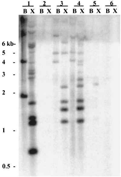
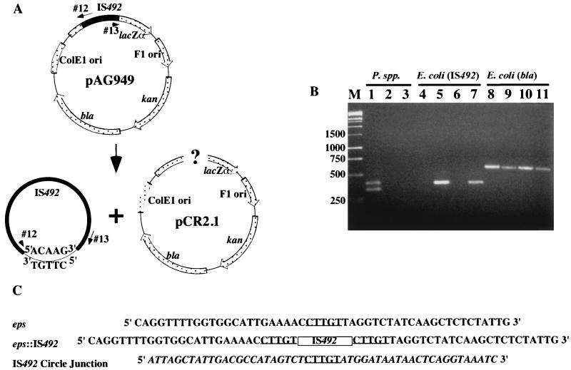
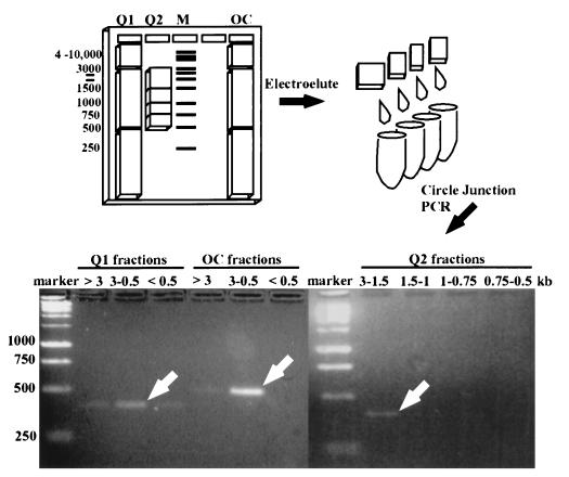
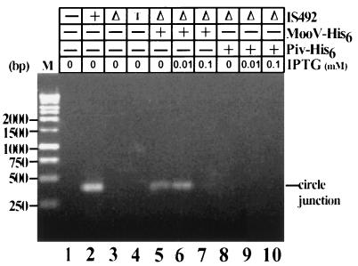
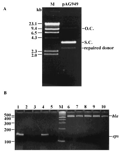
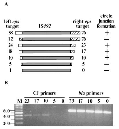
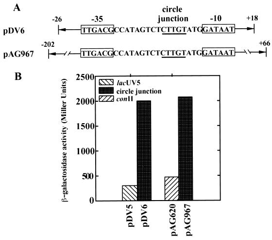
The gram-negative marine bacterium Pseudoalteromonas atlantica produces extracellular polysaccharide (EPS) that is important in biofilm formation by this bacterium. Insertion and precise excision of IS492 at a locus essential for extracellular polysaccharide production (eps) controls phase variation of EPS production in P. atlantica. Examination of IS492 transposition in P. atlantica by using a PCR-based assay revealed a circular form of IS492 that may be an intermediate in transposition or a terminal product of excision. The DNA sequence of the IS492 circle junction indicates that the ends of the element are juxtaposed with a 5-bp spacer sequence. This spacer sequence corresponds to the 5-bp duplication of the chromosomal target sequence found at all IS492 insertion sites on the P. atlantica chromosome that we identified by using inverse PCR. IS492 circle formation correlated with precise excision of IS492 from the P. atlantica eps target sequence when introduced into Escherichia coli on a plasmid. Deletion analyses of the flanking host sequences at the eps insertion site for IS492 demonstrated that the 5-bp duplicated target sequence is essential for precise excision of IS492 and circle formation in E. coli. Excision of IS492 in E. coli also depends on the level of expression of the putative transposase, MooV. A regulatory role for the circular form of IS492 is suggested by the creation of a new strong promoter for expression of mooV by the joining of the ends of the insertion sequence element at the circle junction.


Figures

FIG. 1
FIG. 1
Southern analysis of P. atlantica and closely related marine bacteria with 32P-labeled IS492 DNA as a probe. Chromosomal DNAs from the following strains were digested with either BamHI alone (lanes B) or BamHI and XmnI (lanes X) and subjected to agarose gel electrophoresis: P. atlantica DB27 (lanes 1), P. atlantica ATCC 19262 (lanes 2), P. atlantica ATCC 43666 (lanes 3), P. atlantica ATCC 43667 (lanes 4), P. espejiana ATCC 29659 (lanes 5), and P. haloplanktis ATCC 14393 (lanes 6). After transfer to a nylon membrane, the DNA was probed with a 1-kb [32P]dATP-labeled IS492 DNA fragment. The positions and sizes of the molecular size markers on the agarose gel are indicated to the left.
FIG. 2
FIG. 2
Detection of IS492 circles in vivo by PCR analysis. (A) Diagram of IS492-containing plasmid pAG949 and locations of primers (oligonucleotides 12 and 13) used in this PCR analysis. Potential products of excision are drawn below the diagram. (B) Agarose gel electrophoresis of products generated from colony PCR with the circle junction primers (oligonucleotides 12 and 13) and total DNA from P. atlantica DB27 (lane 1), P. espejiana (lane 2), P. haloplanktis (lane 3), E. coli InvαF′ containing pCR2.1 (lane 4), InvαF′ containing pAG949 (lane 5), E. coli HMS174(DE3) containing pCR2.1 (lane 6), or HMS174(DE3) containing pAG949 (lane 7). Lanes 8 to 11 have the same template DNAs as lanes 4 to 7, respectively, but PCR was performed with the bla primers (see Materials and Methods), which amplify the β-lactamase gene of pAG949, as a control for template addition. Lane M, molecular size markers in base pairs. (C) Sequences of the eps locus prior to and after IS492 insertion and the circle junction sequence with flanking IS492 termini. The 5-bp target duplication-circle junction sequence is underlined, the IS492 insertion in eps is represented by a box, and the IS492 sequence is italicized in the circle junction sequence.
FIG. 3
FIG. 3
Demonstration of circular product from IS492 excision by electrophoretic fractionation of circular DNA from E. coli containing IS492. The illustration shows the 1% agarose gel used to fractionate the open circular IS492 control DNA (OC) and the Qiagen plasmid (circular) DNA preparations (Q1 and Q2) from E. coli containing pAG949, which carries wild-type IS492. Slices were cut from the gel based on comigration with the DNA molecular size standards (1-kb ladder; Promega). DNAs electroeluted from these slices were used as template DNAs for PCR analysis to detect the IS492 circle junction. The PCR products from the wide-range molecular size gel slices (Q1 and OC, >3, 3 to 0.5, and <0.5 kb) and from the narrow-range molecular size slices (Q2, 3 to 1.5, 1.5 to 1, 1 to 0.75, and 0.75 to 0.5 kb) were separated on a 2% agarose gel. The white arrows indicate the primary circle junction PCR product for each group of fractions.
FIG. 4
FIG. 4
Effect of MooV-His6 and Piv-His6 on IS492 circle formation. IS492 circle formation was examined in E. coli HMS174(DE3) by the PCR assay diagrammed in Fig. 1. The circle junction PCR products after agarose gel electrophoresis are shown. MooV-His6 and Piv-His6 (presence or absence is indicated by + or −, respectively) were provided in trans from pAG954 and pAG955, respectively, to the IS492ΔmooV::cam-carrying plasmid, pAG957 (Δ). The levels of MooV-His6 and Piv-His6 were varied by using different concentrations of IPTG for induction. The following plasmids in HMS174(DE3) were included as controls: pCR2.1 (− for IS492), pAG949 (+ for IS492), and pAG903 (T, IS492 target site mutant [see Materials and Methods]). Lane M, Promega 1-kb markers.
FIG. 5
FIG. 5
Detection of the repaired donor DNA after IS492 excision. (A) Plasmid preparation of pAG949 after agarose gel electrophoresis. The following forms of the plasmid are indicated to the right of the gel: open circular (O.C.), supercoiled (S.C.), and the repaired pAG949 after excision of IS492 (repaired donor). The DNA molecular size marker (lane M) is the Promega λ/H3 markers, with sizes indicated to the left. (B) PCR assay for the repaired donor DNA, using primers complementary to eps (lanes 1 to 5), and controls for addition of template to the reaction mixtures, using primers complementary to bla (lanes 6 to 10). Strains tested for the repaired donor DNA were HMS174(DE3) containing pCR2.1 (no IS492) (lanes 5 and 6), pAG949 (wild-type IS492) (lanes 4 and 7), pAG957 (IS492ΔmooV::cam) (lanes 3 and 8), pAG957 and pAG954 (IS492ΔmooV::cam plus mooV-his6) (lanes 2 and 9), and pAG957 and pAG954 (inducing conditions [0.01 mM IPTG]) (lanes 1 and 10). The molecular size marker (lane M) is the New England Biolabs 100-bp marker with sizes (in base pairs) indicated to the left.
FIG. 6
FIG. 6
Target site requirements for IS492 excision. (A) Schematic diagram showing the extent of eps target sequence on either side of IS492 (left flanking sequence, open box; right flanking sequence, hatched box). The number at either end of IS492 indicates the number of nucleotides of eps sequence. Whether a circle junction PCR product could be detected is indicated as + or −. (B) Agarose gel electrophoresis of circle junction (CJ primers) PCR products and bla (bla primers) control products generated from plasmids carrying IS492 sequence with the flanking sequences shown in panel A. Lane M, Promega 1-kb marker, with the sizes of some of the markers (in base pairs) shown to the left.
FIG. 7
FIG. 7
Identification of other target sites in P. atlantica DB27. (A) Agarose gel electrophoresis of PCR products generated from inverse PCR (described in Materials and Methods) of diluted and ligated P. atlantica DB27 chromosomal DNA following digestion with either RsaI (lanes 1 to 3) or HaeIII (lanes 4 to 6). Either 0.25 μg (lanes 1 and 4), 0.025 μg (lanes 2 and 5), or 0.0005 μg (lanes 3 and 6) of P. atlantica chromosomal DNA was used as a template in inverse PCRs. Lane M, Promega 1-kb marker. The inverse image of the UV-illuminated ethidium bromide-stained gel is shown. (B) Alignment of target site sequences. The duplicated target sequence is indicated with a box.
FIG. 8
FIG. 8
Promoter activity associated with the IS492 circle junction sequence. (A) The potential promoter sequence created when IS492 ends are joined at the circle junction is indicated by boxes. Relevant regions of plasmids pDV6 and pAG967, used to evaluate circle junction promoter activity, are schematically shown. Arrows and numbers (base pairs) are used to indicate the region of IS492 included on either side of the circle junction on these plasmids. The circle junction sequence is underlined. (B) Promoter activity measured as β-galactosidase activity (Miller units) in plasmids having constitutive control promoters (lacUV5 [pDV5] or conII [pAG620]) or the circle junction and surrounding IS492 sequence (pDV6 or pAG967). Besides having different amounts of surrounding IS492 sequence, pDV6 and pAG967 have different copy numbers (multiple and single copy, respectively). The relative β-galactosidase activities for these promoter constructions remained constant in multiple experiments.

References

    1. Ausubel F M, Brent R, Kingston R E, Moore D D, Seidman J G, Smith J A, Struhl K, editors. Current protocols in molecular biology. New York, N.Y: Wiley; 1993.
    1. Bartlett D H, Wright M E, Silverman M. Variable expression of extracellular polysaccharide in the marine bacterium Pseudomonas atlantica is controlled by genome rearrangement. Proc Natl Acad Sci USA. 1988;85:3923–3927. - PMC - PubMed
    1. Bartlett D H, Silverman M. Nucleotide sequence of IS492, a novel insertion sequence causing variation in extracellular polysaccharide production in the marine bacterium Pseudomonas atlantica. J Bacteriol. 1989;171:1763–1766. - PMC - PubMed
    1. Belas R, Bartlett D, Silverman M. Cloning and gene replacement mutagenesis of a Pseudomonas atlantica agarase gene. Appl Environ Microbiol. 1988;54:30–37. - PMC - PubMed
    1. Chan K Y, Baumann L, Garza M M, Baumann P. Two new species of Alteromonas: Alteromonas espejiana and Alteromonas undina. Int J Syst Bacteriol. 1978;28:217–222.

Publication types

MeSH terms

LinkOut - more resources