. 1999 Aug;181(16):5017-23.
doi: 10.1128/JB.181.16.5017-5023.1999.

In Thermoanaerobacterium thermosulfurigenes EM1 S-layer homology domains do not attach to peptidoglycan

Affiliations

In Thermoanaerobacterium thermosulfurigenes EM1 S-layer homology domains do not attach to peptidoglycan

E Brechtel et al. J Bacteriol. 1999 Aug.

Abstract

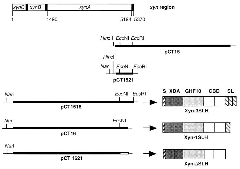
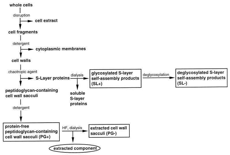
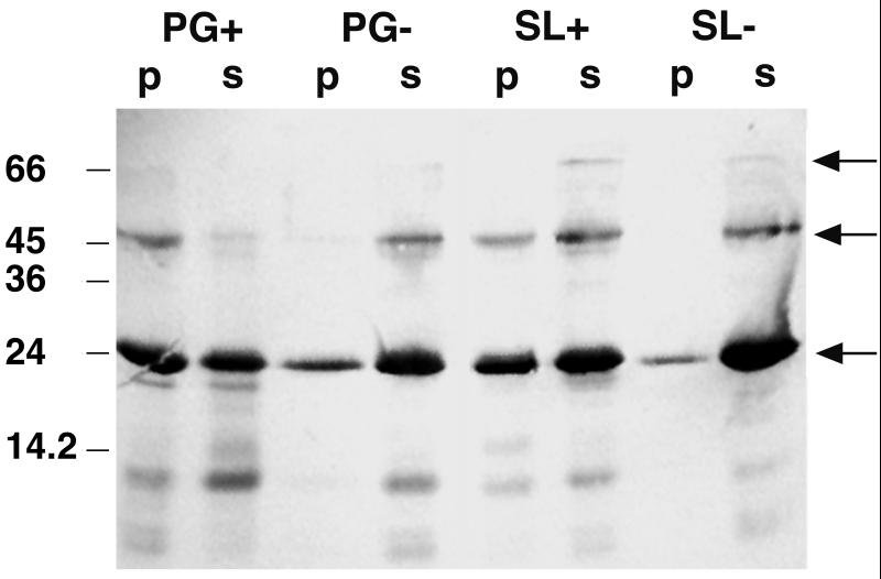
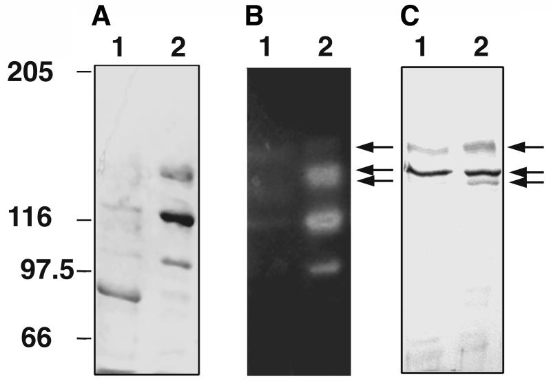
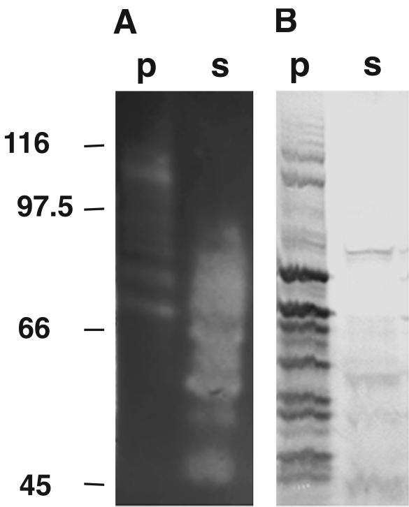
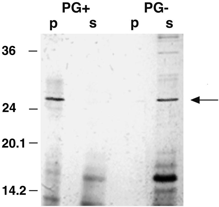
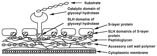
Three exocellular enzymes of Thermoanaerobacterium thermosulfurigenes EM1 possess a C-terminal triplicated sequence related to a domain of bacterial cell surface proteins (S-layer proteins). At least one copy of this sequence, named the SLH (for S-layer homology) domain, is also present at the N terminus of the S-layer protein of this bacterium. The hypothesis that SLH domains serve to anchor proteins to the cell surface was investigated by using the SLH domain-containing xylanase. This enzyme was isolated from T. thermosulfurigenes EM1, and different forms with and without SLH domains were synthesized in Escherichia coli. The interaction of these proteins with isolated components of the cell envelope was determined to identify the attachment site in the cell wall. In addition, a polypeptide consisting of three SLH domains and the N terminus of the S-layer protein of T. thermosulfurigenes EM1 were included in these studies. The results indicate that SLH domains are necessary for the attachment of these proteins to peptidoglycan-containing sacculi. Extraction of the native sacculi with hydrofluoric acid led to the conclusion that not peptidoglycan but accessory cell wall polymers function as the adhesion component in the cell wall. Our results provide further evidence that attachment of proteins via their SLH domains represents an additional mode to display polypeptides on the cell surfaces of bacteria.


Figures

FIG. 1
FIG. 1
Schematic representation of the chromosomal xyn region from T. thermosulfurigenes EM1; DNA fragments cloned on plasmids pCT15, pCT1521, pCT1516, pCT16, and pCT1621; and the domain structures of XynA and XynA derivatives encoded on the plasmids. S, signal peptide; XDA, xylanase domains A; GHF10, catalytic domain of glycosyl hydrolase family 10; CBD, cellulose-binding domains; SL, SLH domains. Restriction sites in pUC18 (thin line) and the inserted gene fragments (thick line) are indicated.
FIG. 2
FIG. 2
Schematic overview of the cell wall preparation procedure and the resulting fractions. Boxed fractions were used in interaction studies. Abbreviations used for the cell wall components are in parentheses. The circled fraction was used for analysis of the extracted component.
FIG. 3
FIG. 3
Western blot showing the affinity of the SLH polypeptide to itself and to isolated cell wall components. Ten micrograms of the SLH polypeptide and 0.5 mg of the cell wall component in a total volume of 100 μl were used in the interaction study. Twenty microliters of each fraction (p and s) was applied to an SDS gel. The blot was prepared with antibodies against the SLH polypeptide. Fractions p and s represent bound and unbound proteins, respectively. Arrows indicate bands representing the SLH polypeptide monomer (23 kDa), dimer, and trimer.
FIG. 4
FIG. 4
Analysis of xylanase purified from the culture supernatant of T. thermosulfurigenes EM1. (A) Coomassie blue-stained SDS gel; (B) activity staining for xylanases; (C) Western blot prepared with antibodies against the SLH domains of the xylanase. Lanes 1, concentrated culture supernatant; lanes 2, concentrated fraction obtained after affinity chromatography. One to 2 μg of protein was applied in each lane. Five different xylanase forms were observed. Arrows indicate xylanase derivatives containing SLH domains. Numbers on the left indicate migrations of marker proteins (in kilodaltons).
FIG. 5
FIG. 5
Interaction of the Xyn-3SLH produced from E. coli(pCT1516) with native peptidoglycan-containing cell wall sacculi (PG+). The xylanase (150 mU) was incubated with 1 mg of the PG+ fraction (total volume, 300 μl), and 20 μl of each sample was used for SDS-PAGE. (A) activity-stained SDS gel; (B) Western blot prepared with antibodies against the SLH polypeptide. Fractions p and s represent bound and unbound proteins, respectively. The migrations of marker proteins (in kilodaltons) are indicated on the left.
FIG. 6
FIG. 6
Interaction of an N-terminal cleavage fragment of the S-layer protein with native peptidoglycan-containing cell wall sacculi (PG+). A silver-stained SDS gel is shown. Fractions containing 20 μg of the cleavage fragments and 0.5 mg of the cell wall components were used (total volume, 100 μl), and 20 μl of each sample was applied to the gel. Bands representing the N-terminal cleavage fragment of the S-layer protein (27 kDa) are indicated by an arrow. p, fraction containing protein bound to the corresponding cell wall component; s, unbound protein. The migrations of marker proteins (in kilodaltons) are indicated on the left.
FIG. 7
FIG. 7
Hypothetic model for the attachment of exocellular proteins from T. thermosulfurigenes EM1 to the cell envelope via their SLH domains. The SLH domains of the S-layer as well as the SLH domains of exocellular glycosyl hydrolases interact with secondary cell wall polymers, which are covalently linked to the underlying peptidoglycan layer.

References

    1. Antranikian G. Physiology and enzymology of thermophilic anaerobic bacteria degrading starch. FEMS Microbiol Rev. 1990;75:201–218.
    1. Antranikian G, Herzberg C, Gottschalk G. Production of thermostable α-amylase, pullulanase, and α-glucosidase in continuous culture by a new Clostridium isolate. Appl Environ Microbiol. 1987;53:1668–1673. - PMC - PubMed
    1. Bergmeyer H U, Gawehn K, Grabe M. Enzyme als biochemische Reagenzien. In: Bergmeyer H U, editor. Methoden der enzymatischen Analyse. Weinheim, Germany: Verlag Chemie; 1974. pp. 454–558.
    1. Birnboim H C, Doly J. A rapid alkaline extraction procedure for screening recombinant plasmid DNA. Nucleic Acids Res. 1979;7:1513–1523. - PMC - PubMed
    1. Blum H, Beier H, Gross H J. Improved silver staining method of plant proteins, RNA and DNA in polyacrylamide gels. Electrophoresis. 1987;8:93–99.

Publication types

MeSH terms

LinkOut - more resources