. 1999 Aug;181(16):5024-32.
doi: 10.1128/JB.181.16.5024-5032.1999.

The bvr locus of Listeria monocytogenes mediates virulence gene repression by beta-glucosides

Affiliations

The bvr locus of Listeria monocytogenes mediates virulence gene repression by beta-glucosides

K Brehm et al. J Bacteriol. 1999 Aug.

Abstract

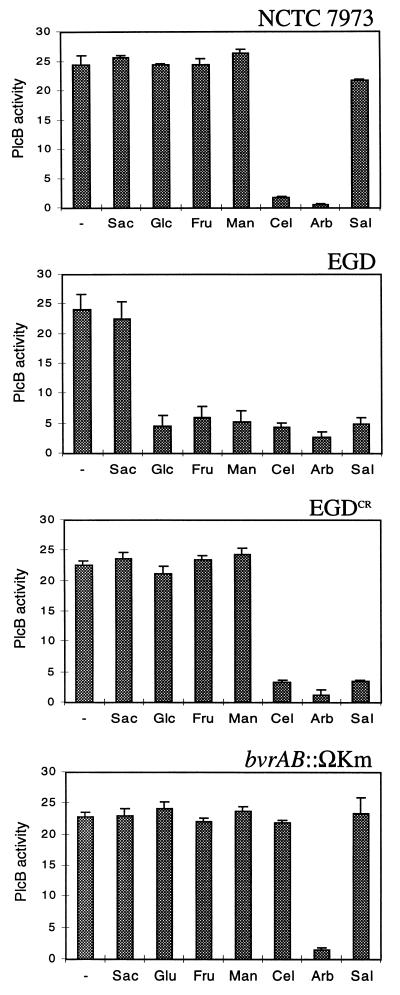
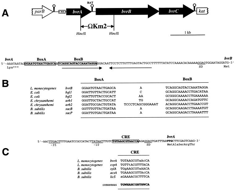
The beta-glucoside cellobiose has been reported to specifically repress the PrfA-dependent virulence genes hly and plcA in Listeria monocytogenes NCTC 7973. This led to the hypothesis that beta-glucosides, sugars of plant origin, may act as signal molecules, preventing the expression of virulence genes if L. monocytogenes is living in its natural habitat (soil). In three other laboratory strains (EGD, L028, and 10403S), however, the effect of cellobiose was not unique, and all fermentable carbohydrates repressed hly. This suggested that the downregulation of virulence genes by beta-glucosides is not a specific phenomenon but, rather, an aspect of a global regulatory mechanism of catabolite repression (CR). We assessed the effect of carbohydrates on virulence gene expression in a panel of wild-type isolates of L. monocytogenes by using the PrfA-dependent phospholipase C gene plcB as a reporter. Utilization of any fermentable sugar caused plcB repression in wild-type L. monocytogenes. However, an EGD variant was identified in which, as in NCTC 7973, plcB was only repressed by beta-glucosides. Thus, the regulation of L. monocytogenes virulence genes by sugars appears to be mediated by two separate mechanisms, one presumably involving a CR pathway and another specifically responding to beta-glucosides. We have identified in L. monocytogenes a 4-kb operon, bvrABC, encoding an antiterminator of the BglG family (bvrA), a beta-glucoside-specific enzyme II permease component of the phosphoenolpyruvate-sugar phosphotransferase system (bvrB), and a putative ADP-ribosylglycohydrolase (bvrC). Low-stringency Southern blots showed that this locus is absent from other Listeria spp. Transcription of bvrB was induced by cellobiose and salicin but not by arbutin. Disruption of the bvr operon by replacing part of bvrAB with an interposon abolished the repression by cellobiose and salicin but not that by arbutin. Our data indicate that the bvr locus encodes a beta-glucoside-specific sensor that mediates virulence gene repression upon detection of cellobiose and salicin. Bvr is the first sensory system found in L. monocytogenes that is involved in environmental regulation of virulence genes.


Figures

FIG. 1
FIG. 1
Patterns of virulence gene (plcB) repression in response to sugars in L. monocytogenes strains NCTC 7973, EGD, and EGDCR and the bvrAB::Km mutant (Sac, saccharose; Glc, glucose; Fru, fructose; Man, mannose; Cel, cellobiose; Arb, arbutin; Sal, salicin; –, no sugar). Means for three independent experiments ± the standard error are shown.
FIG. 2
FIG. 2
(A) Scheme of the genetic structure of the bvr locus of L. monocytogenes. The positions of the putative transcriptional terminators, CRE, and RAT sequences are shown. The bvr mutation was created by allele exchange by using a plasmid construct in which a HincII fragment encompassing the 3′-terminal third of bvrA and the 5′-proximal third of bvrB was replaced by the ΩKm2 interposon, as indicated. (B) Nucleotide sequence of the intergenic region between bvrA and bvrB (the respective last and first codons are shown) and position of the putative RAT sequence (boxed), which overlaps the 5′ part of a palindromic structure that may act as transcriptional terminator (indicated by inverted arrows). Below, comparison of the putative RAT sequence preceding bvrB with known RAT sequences (13). (C) Nucleotide sequence of the 3′ region upstream from bvrA (its TTG start codon is in boldface type) showing −10 and −35 putative promoter sequences and the CRE-like sequence (boxed). Below, comparison of the putative CRE preceding bvrA with the CRE-like element of the ccpA promoter region of L. monocytogenes (3) and known active CRE sites from gram-positive bacteria (21). Deviations from the CRE consensus sequence (in boldface) are shown in lowercase (N = any nucleotide; W = A or T).
FIG. 3
FIG. 3
Multiple alignments of the deduced sequences of the polypeptides encoded by the bvr locus with their corresponding protein homologs from various bacteria (A.br, Azospirillum brasilense; B.su, B. subtilis; E.ch, E. chrysanthemi; E.co, E. coli; L.mo., L. monocytogenes; M.ja, M. jannaschii; R.ca, Rhodobacter capsulatus). The Bvr primary structures shown are those predicted from DNA sequences determined from EGDCR (accession number of the bvr locus: AG007877). (A) Alignment of BvrA with known ATs (accession numbers: ArbG, P26211; BglG, P11989; LicT, P39805; SacY, P15401; and SacT, P26212; identities with BvrA: 33, 28, 35, 30, and 29%, respectively). (B) Alignment of BvrB with known β-glucoside-specific enzyme II PTS permease components (accession numbers: ArbF, P26207; BglF, P08722; and BglP, P40739; identities with BvrB: 37, 34, and 32%, respectively). The positions of residues involved in the catalytic function of the E. coli BglF permease, as determined by site-specific mutagenesis (49), are indicated by black symbols: inverted triangles, Cys-24 and His-306 residues essential for the transfer of the phosphoryl group to the sugar; stars, His-547 and Asp-551 residues involved in the phosphorylation of the permease by HPr; and circle, His-183 residue important for substrate specificity. Note that almost all of these catalytically relevant residues are conserved in BvrB and related permeases. The only exception is the histidyl residue that aligns with position 183 of BglF, which is absent from the BvrB sequence. This sequence divergence may account for the differences in substrate specificity between BvrB and other β-glucoside permeases (see the text). (C) Alignment of BvrC with DraG proteins from R. capsulatus (accession number X71131) and A. brasilense (accession number I39752) and DraG homologs encoded in the genomes of M. jannaschii (accession number C64448) and E. coli (hypothetical protein b2099; accession number B64977). In the nitrogen-fixing bacteria R. rubrum, A. brasiliense, and R. capsulatus, dinitrogenase reductase (an enzyme essential for nitrogen fixation) is inhibited by ADP-ribosylation catalyzed by DraT (dinitrogenase reductase ADP-ribosyltransferase) and activated by the removal of the ADP-ribosyl group catalyzed by the ADP-ribosylglycohydrolase DraG (dinitrogenase reductase activating glycohydrolase) (30, 37, 56). Posttranslational modification via ADP-ribosylation has also been shown to be important in the regulation of glutamine synthetase in R. rubrum and Rhizobium meliloti and eventually also of sporulation in B. subtilis (22).
FIG. 4
FIG. 4
RT-PCR transcription analysis of bvrB (A) and plcB (B) expression in EGDCR grown in the presence of cellobiose (Cel), salicin (Sal), arbutin (Arb), and glucose (Glc) (control). Note that in panel A, cellobiose and salicin, but not arbutin, upregulate bvrB. In panel B, the three β-glucosides repress plcB, a finding consistent with the data obtained by using PlcB activity as reporter (Fig. 1).
FIG. 5
FIG. 5
Detection of bvr sequences in Listeria spp. by low-stringency Southern blot. Lanes: a, L. monocytogenes EGDCR; b, L. monocytogenes NCTC 7973; c, L. ivanovii ATCC 19119; d, L. seeligeri SLCC 5921; e, L. welshimeri SLCC 5334; f, L. grayi. Chromosomal DNA was digested with EcoRI. Numbers on the left indicate the size in kilobases.

References

    1. Ausubel F M, Brent R, Kingston R E, Moore D D, Seidman J G, Smith J A, Struhl K. Current protocols in molecular biology. New York, N.Y: John Wiley & Sons; 1991.
    1. Behari J, Youngman P. Regulation of hly expression in Listeria monocytogenes by carbon sources and pH occurs through separate mechanisms mediated by PrfA. Infect Immun. 1998;66:3635–3642. - PMC - PubMed
    1. Behari J, Youngman P. A homolog of CcpA mediates catabolite control in Listeria monocytogenes but not carbon source regulation of virulence genes. J Bacteriol. 1998;180:6316–6324. - PMC - PubMed
    1. Bohne J, Sokolovic Z, Goebel W. Transcriptional regulation of prfA and PrfA-regulated virulence genes in Listeria monocytogenes. Mol Microbiol. 1994;11:1141–1150. - PubMed
    1. Brehm K, Haas A, Goebel W, Kreft J. A gene encoding a superoxide dismutase of the facultative intracellular bacterium Listeria monocytogenes. Gene. 1992;118:121–125. - PubMed

Publication types

MeSH terms

Associated data

LinkOut - more resources