

doi: 10.1128/JB.181.16.5114-5118.1999.
In vivo observation of cell division of anaerobic hyperthermophiles by using a high-intensity dark-field microscope
Affiliations
- PMID: 10438790
- PMCID: PMC94007
- DOI: 10.1128/JB.181.16.5114-5118.1999
Item in Clipboard
In vivo observation of cell division of anaerobic hyperthermophiles by using a high-intensity dark-field microscope
J Bacteriol.
1999 Aug.
Abstract
To study growth and cell division of anaerobic hyperthermophilic archaea in vivo, a cultivation technique using glass capillaries was developed. At temperatures of 90 to 98 degrees C, at least 10 successive cell divisions of Pyrodictium abyssi TAG 11 were documented. Cells divide by binary fission. Visualized under a modified dark-field microscope, the formation of cannulae, which finally connected all cells, was observed. The cannulae elongated at 1.0 to 1.5 micrometers/min and reached final lengths of between 30 and 150 micrometers. A "snapping division"-like mode of cell fission was discovered for Thermoproteus tenax.
Figures
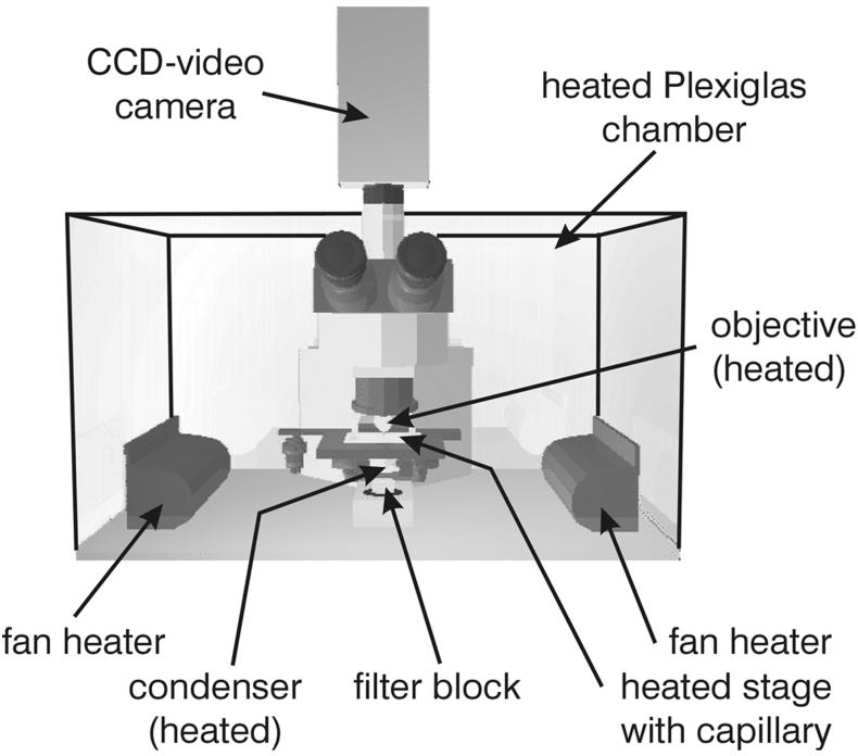
Scheme of the microscope including heatable polyacrylate chamber and video camera. CCD, charge-coupled device.
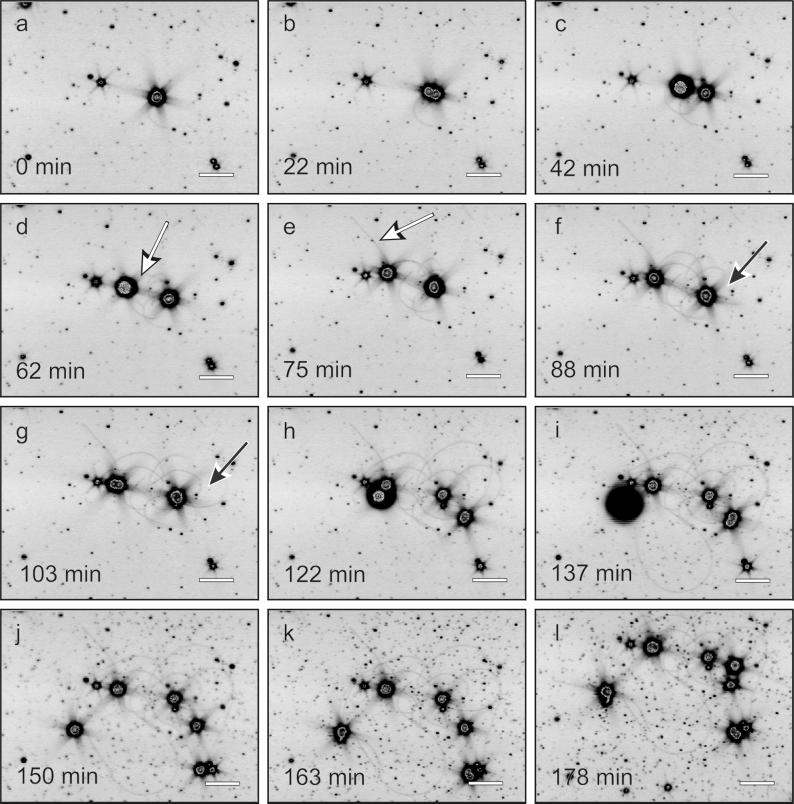
Cell division of P. abyssi TAG 11 and growth of cannulae. Frames were extracted from interval recording; for details, see the text. White arrows indicate cannulae with one insertion point and one free end; black arrows indicate cannula loops. Scale bar, 10 μm.
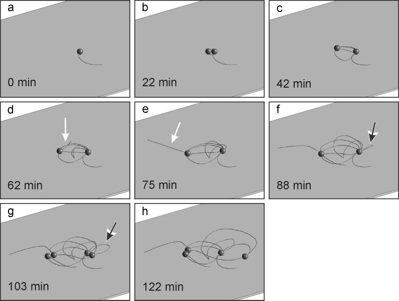
Two-dimensional reconstruction of cell division of P. abyssi TAG 11 and development of the network. Frames a to h were calculated from the data of Fig. 2a to 2h. White arrows indicate cannulae with one insertion point and one free end; black arrows indicate cannula loops.

Cell division of T. tenax. Frames were obtained from time-lapse film. Scale bar, 20 μm.
References
-
- Hotani H, Asakura S. Growth-saturation in vitro of Salmonella flagella. J Mol Biol. 1974;86:285–300. - PubMed
-
- Inoue S. The role of microtubule assembly dynamics in mitotic force generation and functional organization of living cells. J Struct Biol. 1986;118:87–93. - PubMed
-
- König H, Messner P, Stetter K O. The fine structure of the fibers of Pyrodictium occultum. FEMS Microbiol Lett. 1988;49:207–212.
-
- Lotz G. Verzeichnis der Wissenschaftlichen Filme. Biologie. Göttingen, Germany: IWF; 1986.
Publication types
MeSH terms
LinkOut - more resources
Full Text Sources
