. 1999 Sep;73(9):7153-64.
doi: 10.1128/JVI.73.9.7153-7164.1999.

The herpes simplex virus vhs protein induces endoribonucleolytic cleavage of target RNAs in cell extracts

Affiliations

The herpes simplex virus vhs protein induces endoribonucleolytic cleavage of target RNAs in cell extracts

M M Elgadi et al. J Virol. 1999 Sep.

Abstract

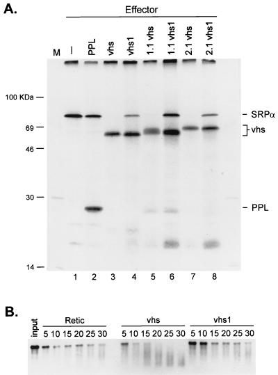
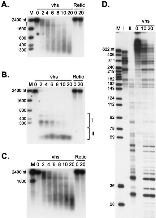
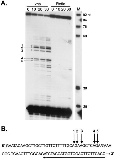
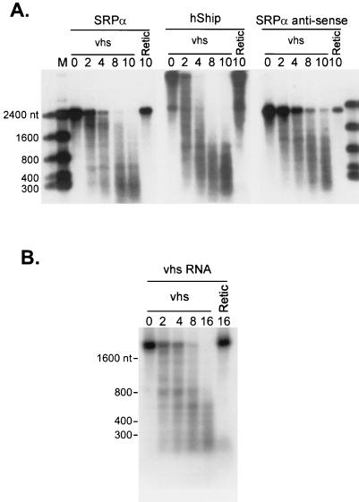
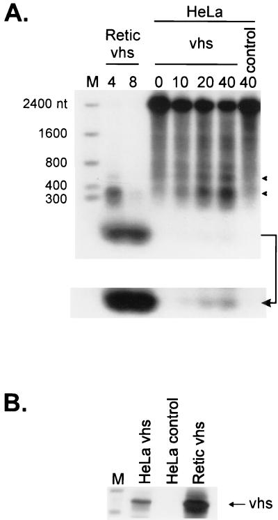
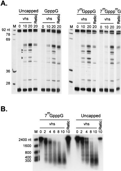
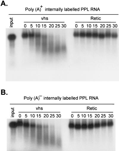
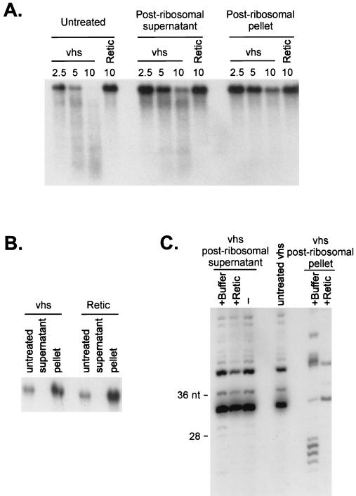
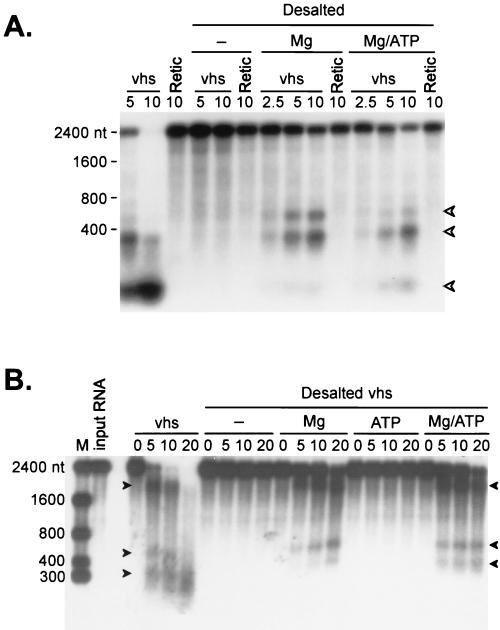
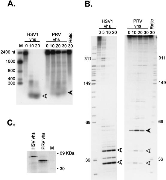
The herpes simplex virus virion host shutoff (vhs) protein (UL41 gene product) is a component of the HSV virion tegument that triggers shutoff of host protein synthesis and accelerated mRNA degradation during the early stages of HSV infection. Previous studies have demonstrated that extracts from HSV-infected cells and partially purified HSV virions display vhs-dependent RNase activity and that vhs is sufficient to trigger accelerated RNA degradation when expressed as the only HSV protein in an in vitro translation system derived from rabbit reticulocytes. We have used the rabbit reticulocyte translation system to characterize the mode of vhs-induced RNA decay in more detail. We report here that vhs-dependent RNA decay proceeds through endoribonucleolytic cleavage, is not affected by the presence of a 5' cap or a 3' poly(A) tail in the RNA substrate, requires Mg(2+), and occurs in the absence of ribosomes. Intriguingly, sites of preferential initial cleavage were clustered over the 5' quadrant of one RNA substrate that was characterized in detail. The vhs homologue of pseudorabies virus also induced accelerated RNA decay in this in vitro system.


Figures

FIG. 1
FIG. 1
HSV-1 vhs induces translational arrest and mRNA degradation in vitro. (A) RRL were programmed with the indicator effector mRNAs, and translation was allowed to proceed for 20 mins in the presence of [35S]methionine. Lysates were then challenged with an equal amount of capped SRPα reporter mRNA, and the reactions were allowed to continue for an additional 60 min. The translation products were resolved on an SDS–12% polyacrylamide gel, and the 35S signal was detected by autoradiography with Kodak X-Omat AR film. 1.1 and 2.1, doubly tagged active vhs variants; vhs1, 1.1 vhs1, and 2.1 vhs1, inactive vhs point mutant derivatives of vhs, 1.1, and 2.1. (B) RRL were programmed with vhs RNA (lanes vhs), vhs1 RNA (lanes vhs1), or no RNA (control, lanes Retic), and translation was allowed to proceed for 20 min. The lysates were then challenged with capped, internally labeled SRPα mRNA. Samples were recovered at the indicated times (numbers above lanes, in minutes), and the RNA reaction products were resolved on a 1% agarose–6% formaldehyde gel, transferred to a Nytran Plus membrane, and detected by autoradiography with Kodak X-Omat AR film.
FIG. 2
FIG. 2
Analysis of vhs-induced degradation intermediates of SRPα RNA. (A and B) Internally labeled (A) and cap-labeled (B) SRPα mRNAs were added to RRL containing pretranslated vhs (lanes vhs) or RRL control (lanes Retic). RNA degradation products were recovered at the indicated times (minutes) and analyzed by agarose-formaldehyde gel electrophoresis. (C) The membrane in panel B was hybridized to a 32P-labeled DNA probe corresponding to the 3′-most 400 nt of SRPα RNA (after the radioactive signal from the cap label had been allowed to decay for six half-lives). The bound probe was then detected by autoradiography with Kodak X-Omat AR film. Numbers to the left of panels A, B, and C represent the sizes of RNA markers (lanes M) in nucleotides. (D) Cap-labeled SRPα RNA was added to RRL-vhs, and the RNA degradation intermediates recovered at 10 min were resolved on a 1% agarose–6% formaldehyde gel. RNA fragments contained in the gel slices indicated by brackets I and II in panel C were eluted and resolved on an 8% polyacrylamide sequencing gel (lanes I and II) along with the unfractionated products of a vhs reaction on cap-labeled SRPα RNA (sampled at 0, 10, and 20 min). Numbers to the left of panel D represent the sizes of DNA markers (lane M) in nucleotides.
FIG. 3
FIG. 3
Primer extension analysis of the 5′-most degradation products of SRPα RNA. (A) Unlabeled, capped SRPα RNA was added to RRL-vhs (lanes vhs) or RRL control (lanes Retic), and RNA reaction products were recovered at the indicated time points (minutes). The RNA reaction products were then analyzed by primer extension with 5′-32P-labeled oligonucleotide complementary to nt 60 to 84 of the SRPα RNA. Primer extension products were resolved on an 8% polyacrylamide sequencing gel and detected by autoradiography. Numbered arrows indicate the positions of primer extension products representing vhs-induced novel 5′ ends. Numbers to the right of lane M (marker) indicate the sizes of DNA markers in nucleotides. (B) Sequence of the extreme 5′ 84 nt of SRPα RNA. Numbered arrows above the sequence correspond to those in panel A and indicate the positions of vhs-induced cleavage at the 5′ end of SRPα RNA. The arrow under the sequence indicates the position of the oligonucleotide primer used.
FIG. 4
FIG. 4
vhs induces degradation of a variety of RNA substrates. (A) Internally labeled SRPα (2.4 kb), hSHIP (4.5 kb), and SRPα antisense (2.2 kb) RNAs were added to RRL containing vhs (lanes vhs) or RRL control (lanes Retic), and samples recovered at the indicated times (minutes) were analyzed by agarose-formaldehyde gel electrophoresis as in Fig. 1B. (B) Internally labeled vhs RNA (1.8 kb) was reacted with RRL containing vhs or control RRL, and samples recovered at the indicated times (minutes) were analyzed as in panel A.
FIG. 5
FIG. 5
vhs synthesized in a HeLa cell translation extract induces degradation of SRPα RNA. (A) HeLa cell translation extracts and RRL were programmed with capped unlabeled vhs RNA, and translation was allowed to proceed for 60 min in the presence of [35S]methionine. Extracts were then challenged with cap-labeled SRPα RNA, and samples were recovered at the indicated times (minutes). Reaction products were then analyzed by agarose-formaldehyde gel electrophoresis as in Fig. 1B. The bottom of panel A shows an overexposure of the lower portion of the membrane in panel A (indicated by an arrow). Lane control, HeLa cell extracts lacking vhs RNA. Numbers to the left of panel A indicate the sizes of RNA markers (lane M) in nucleotides. Solid arrowheads indicate the positions of vhs-induced RNA degradation products. (B) Samples of the translation reaction products used in panel A were resolved on an SDS–12% polyacrylamide gel, and the 35S signal was detected by autoradiography.
FIG. 6
FIG. 6
vhs-induced RNA degradation is cap independent. (A) Unlabeled SRPα RNAs bearing the indicated 5′ cap structures were added to RRL-vhs (lanes vhs) and control RRL (lanes Retic), and samples were recovered at the indicated times (minutes). The RNA reaction products were then analyzed by primer extension with 5′ 32P-labeled oligonucleotide complementary to residues 60 to 84 of the SRPα RNA, as in Fig. 3. Solid arrowheads indicate the mobilities of the vhs-induced products. Numbers to the left of panel A indicate the sizes of DNA markers (lanes M) in nucleotides. (B) Internally labeled capped (7mGpppG) and uncapped SRPα RNAs were added to RRL containing vhs (lanes vhs) and RRL control (lanes Retic). Samples recovered at the indicated times (minutes) were then analyzed by agarose-formaldehyde gel electrophoresis. Numbers to the left of panel B indicate the sizes of RNA markers (lane M) in nucleotides.
FIG. 7
FIG. 7
vhs-induced RNA degradation is not influenced by a 3′ poly(A) tail. Internally labeled PPL RNA containing (A) or lacking (B) a ∼35-residue poly(A) tail followed by GU was incubated with RRL vhs (lanes vhs) and RRL control (lanes Retic) for the indicated times (minutes). RNA reaction products were then analyzed by agarose-formaldehyde gel electrophoresis as in Fig. 1. Lanes 1. input, untreated RNAs.
FIG. 8
FIG. 8
vhs-induced RNA degradation does not require ribosomes. RRL containing (vhs) or lacking (Retic) vhs were centrifuged at 160,000 × g for 50 min at 4°C to pellet the ribosomes. The ribosomal pellet was resuspended in Retic buffer (1.6 mM Tris acetate [pH 7.8], 80 mM potassium acetate, 2 mM magnesium acetate, 0.25 mM ATP, 0.1 mM DTT). (A) Untreated lysates, postribosomal supernatants, and pellets were mixed with internally labeled SRPα RNA, and samples recovered at various times (minutes) were analyzed by agarose-formaldehyde gel electrophoresis as in Fig. 1B. (B) A Northern blot analysis of the postribosomal supernatant and pellet fractions was performed with a rabbit 18S rRNA-specific 5′-32P-labeled oligonucleotide probe. (C) Cap-labeled SRPα RNA was added to untreated RRL vhs and to postribosomal supernatants and pellets that had been mixed with an equal volume of Retic buffer (+buffer) or naive RRL (+Retic). RNA samples were recovered after 10 min and then resolved on an 8% polyacrylamide sequencing gel.
FIG. 9
FIG. 9
vhs-induced RNA degradation requires magnesium. RRL containing (vhs) or lacking (Retic) vhs were desalted on Sephadex G-25 spin columns at 4 ml of packed resin per 100 μl of lysate. The resin was swollen in Retic buffer lacking magnesium acetate and ATP, loaded in a glass wool-plugged 5-ml syringe, and precentrifuged for 5 min at 4°C and 1,750 × g in a clinical centrifuge equipped with a swinging-bucket rotor. Samples of the desalted lysates were then combined with and equal volume of Retic buffer containing 4 mM magnesium acetate (lanes Mg), 0.5 mM ATP (lanes ATP) or 4 mM magnesium acetate and 0.5 mM ATP (lanes Mg/ATP). Substrate SRPα RNA was then added, and samples withdrawn at the indicated times (minutes) were analyzed by formaldehyde-agarose gel electrophoresis. (A) Analysis of cap-labeled SRPα RNA. (B) Analysis of internally labeled SRPα RNAs. Arrowheads indicate the mobilities of some of the vhs-dependent RNA degradation intermediates. Numbers to the left of the panels indicate the sizes of RNA markers (lanes M) in nucleotides.
FIG. 10
FIG. 10
PrV vhs displays RNA degradation activity in vitro. RRL were programmed with RNAs encoding HSV1 or PrV vhs, and translation was allowed to proceed for 60 min. Lysates were then challenged with cap-labeled SRPα RNA, and samples recovered at the indicated times (minutes) were analyzed by electrophoresis through an agarose-formaldehyde gel (A) and an 8% polyacrylamide sequencing gel (B). Numbers to the sides of the panels indicate the sizes of marker fragments in nucleotides (lanes M; RNA and DNA in panels A and B, respectively). Solid and open arrowheads indicate the positions of corresponding RNA fragments in the two different gel systems. (C) SDS-polyacrylamide gel electrophoresis of the HSV and PrV vhs proteins produced in the translation reactions in panels A and B.

References

    1. Beelman C A, Parker R. Degradation of mRNA in eukaryotes. Cell. 1995;81:179–183. - PubMed
    1. Ben-Porat T, Rakusanova T, Kaplan A S. Early functions of the genome of herpesvirus. II. Inhibition of formation of cell-specific polysomes. Virology. 1971;46:890–899. - PubMed
    1. Berthomme H, Jacquemont B, Epstein A. The pseudorabies virus host-shutoff homolog gene: nucleotide sequence and comparison with alphaherpesvirus protein counterparts. Virology. 1993;193:1028–1032. - PubMed
    1. Binder R, Horowitz J A, Basilion J P, Koeller D M, Klausner R D, Harford J B. Evidence that the pathway of transferrin receptor mRNA degradation involves an endonucleolytic cleavage within the 3′ UTR and does not involve poly(A) tail shortening. EMBO J. 1994;13:1969–1980. - PMC - PubMed
    1. Brown B D, Zipkin I D, Harland R M. Sequence-specific endonucleolytic cleavage and protection of mRNA in Xenopus and Drosophila. Genes Dev. 1993;7:1620–1631. - PubMed

Publication types

LinkOut - more resources