. 1999 Sep;73(9):7671-7.
doi: 10.1128/JVI.73.9.7671-7677.1999.

An inducible human immunodeficiency virus type 1 (HIV-1) vector which effectively suppresses HIV-1 replication

Affiliations

An inducible human immunodeficiency virus type 1 (HIV-1) vector which effectively suppresses HIV-1 replication

D S An et al. J Virol. 1999 Sep.

Abstract

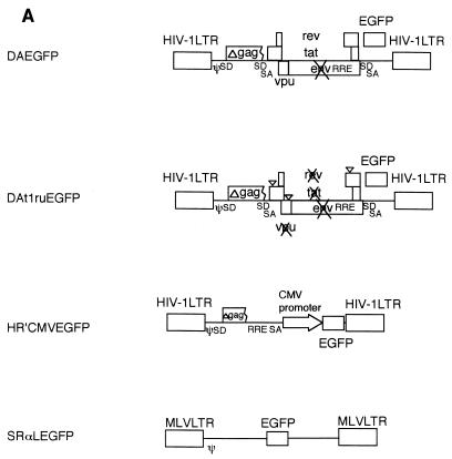
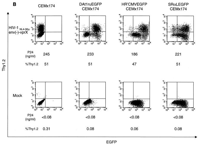
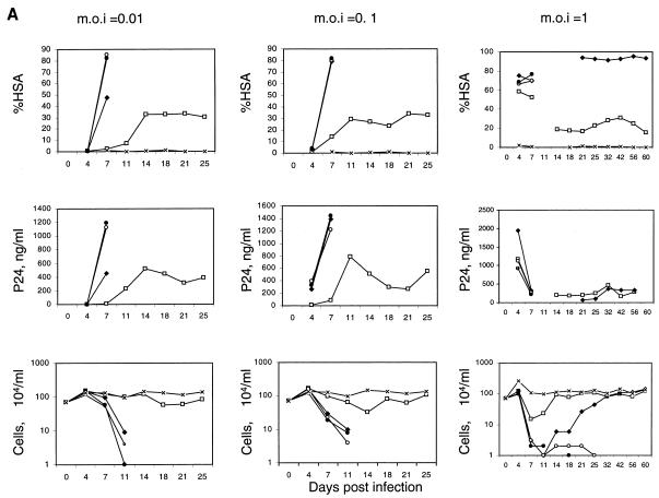
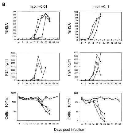
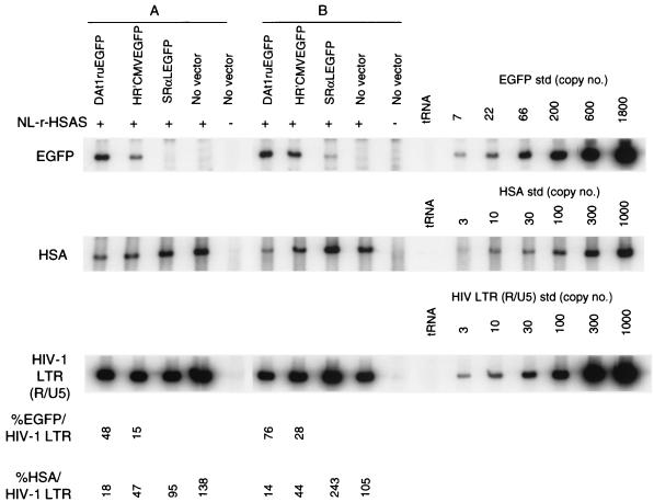
Recently, gene therapy vectors based upon the human immunodeficiency virus type 1 (HIV-1) genome have been developed. Here, we create an HIV-1 vector which is defective for all HIV-1 genes, but which maintains cis-acting elements required for efficient packaging, infection, and expression. In T cells transduced by this vector, vector expression is low but efficiently induced following HIV-1 infection. Remarkably, although the HIV-1 vector does not contain specific anti-HIV-1 therapeutic genes, the presence of the vector alone is sufficient to inhibit the spread of HIV-1 infection. The mechanism of inhibition is likely to be at the level of competition for limiting substrates required for either efficient packaging or reverse transcription, thereby selecting against propagation of wild-type HIV-1. These results provide proof of a concept for potential application of a novel HIV-1 vector in HIV-1 disease.


Figures

FIG. 1
FIG. 1
(A) Maps of vectors. An HIV-1 vector, DAEGFP, contains sequences from HIV-1 NL4-3 but lacks the gag, pol, env, vif, vpr, and nef genes, as described in Materials and Methods. The EGFP gene was cloned in place of the nef gene, and is expressed from HIV-1 LTR. The tat and rev genes are intact for gene expression from HIV-1 LTR. An inducible HIV-1 vector, DAt1ruEGFP, was constructed from DAEGFP by ablation of the tat, rev, and vpu genes, as described in Materials and Methods. All genes from HIV-1 were ablated in the DAt1ruEGFP vector. An HIV-1 vector, pHR′CMVEGFP, lacks all HIV-1 genes and expresses EGFP from the CMV internal promoter, as described in Materials and Methods. A murine retrovirus vector, SRαLEGFP, express EGFP from the MLV LTR, as described in Materials and Methods. The LTR, splice donor and acceptor sites (SD and SA, respectively), the packaging signal (ψ), the truncated gag sequence (Δgag), the frameshift mutation (∇), the rev-responsive element (RRE), CMV, and MLV are indicated. (B) Induction of gene expression from HIV-1 vectors by single-round infection with infectious HIV-1 reporter virus. Mock-transduced (CEMx174) and vector-transduced (DAt1ruEGFP CEMx174, HR′CMVEGFP CEMx174, and SRαLEGFP CEMx174) CEMx174 cells (2 × 10⁵) were infected with VSVG-pseudotyped HIV-1NL4-3thyenv(−)-vprX at an MOI of 0.5 (upper panels) or were mock infected (lower panels). At 3 days postinfection, cells (2 × 10⁵) were stained with a monoclonal antibody to murine Thy1.2 conjugated with PE, and 5 × 10³ cells were analyzed by flow cytometry for EGFP and Thy1.2 expression. The x axis indicates EGFP fluorescence intensity; the y axis indicates Thy1.2 expression. As indicated, p24 production in cell supernatant was also measured by ELISA at 3 days postinfection. %Thy1.2 indicates Thy1.2-positive populations.
FIG. 1
FIG. 1
(A) Maps of vectors. An HIV-1 vector, DAEGFP, contains sequences from HIV-1 NL4-3 but lacks the gag, pol, env, vif, vpr, and nef genes, as described in Materials and Methods. The EGFP gene was cloned in place of the nef gene, and is expressed from HIV-1 LTR. The tat and rev genes are intact for gene expression from HIV-1 LTR. An inducible HIV-1 vector, DAt1ruEGFP, was constructed from DAEGFP by ablation of the tat, rev, and vpu genes, as described in Materials and Methods. All genes from HIV-1 were ablated in the DAt1ruEGFP vector. An HIV-1 vector, pHR′CMVEGFP, lacks all HIV-1 genes and expresses EGFP from the CMV internal promoter, as described in Materials and Methods. A murine retrovirus vector, SRαLEGFP, express EGFP from the MLV LTR, as described in Materials and Methods. The LTR, splice donor and acceptor sites (SD and SA, respectively), the packaging signal (ψ), the truncated gag sequence (Δgag), the frameshift mutation (∇), the rev-responsive element (RRE), CMV, and MLV are indicated. (B) Induction of gene expression from HIV-1 vectors by single-round infection with infectious HIV-1 reporter virus. Mock-transduced (CEMx174) and vector-transduced (DAt1ruEGFP CEMx174, HR′CMVEGFP CEMx174, and SRαLEGFP CEMx174) CEMx174 cells (2 × 10⁵) were infected with VSVG-pseudotyped HIV-1NL4-3thyenv(−)-vprX at an MOI of 0.5 (upper panels) or were mock infected (lower panels). At 3 days postinfection, cells (2 × 10⁵) were stained with a monoclonal antibody to murine Thy1.2 conjugated with PE, and 5 × 10³ cells were analyzed by flow cytometry for EGFP and Thy1.2 expression. The x axis indicates EGFP fluorescence intensity; the y axis indicates Thy1.2 expression. As indicated, p24 production in cell supernatant was also measured by ELISA at 3 days postinfection. %Thy1.2 indicates Thy1.2-positive populations.
FIG. 2
FIG. 2
Kinetics of HSA expression, p24 production, and cell count after infection of replication-competent HIV-1 reporter virus. (A) Mock-transduced and vector-transduced CEMx174 cells (DAt1ruEGFP CEMx174, HR′CMVEGFP CEMx174, and SRαLEGFP CEMx174) (7 × 10⁵) were infected with replication-competent NL-r-HSAS at MOI of 0.01, 0.1, and 1. Every 3 or 4 days, the cultures were divided into fifths and recultured in fresh medium. The remaining four-fifths was analyzed for HSA expression by flow cytometry (%HSA), the amount of p24 in the supernatant was measured by ELISA (p24, ng/ml), and the cells were counted (104), as described in Materials and Methods. Due to the low number of cells in cultures at day 11 postinfection at an MOI of 1, analyses of HSA expression and p24 were not performed at that time point. □, DAt1ruEGFP CEMx174 cells infected with NL-r-HSAS; ⧫, HR′CMVEGFP CEMx174 cells infected with NL-r-HSAS; ○, SRαLEGFP CEMx174 cells infected with NL-r-HSAS; ●, CEMx174 cells infected with NL-r-HSAS virus; ×, CEMx174 cells with no NL-r-HSAS infection. (B) Mock-transduced and vector-transduced SupT1 cells (7 × 10⁵) (DAt1ruEGFP SupT1, HR′CMVEGFP SupT1, SRαLEGFP SupT1) were infected with replication-competent NL-r-HSAS at MOI of 0.01 and 0.1. The cultures were analyzed as described for panel A. □, DAt1ruEGFP SupT1 cells infected with NL-r-HSAS; ⧫, HR′CMVEGFP SupT1 cells infected with NL-r-HSAS; ○, SRαLEGFP SupT1 cells infected with NL-r-HSAS; ●, SupT1 cells infected with NL-r-HSAS virus; ×, SupT1 cells with no NL-r-HSAS infection.
FIG. 2
FIG. 2
Kinetics of HSA expression, p24 production, and cell count after infection of replication-competent HIV-1 reporter virus. (A) Mock-transduced and vector-transduced CEMx174 cells (DAt1ruEGFP CEMx174, HR′CMVEGFP CEMx174, and SRαLEGFP CEMx174) (7 × 10⁵) were infected with replication-competent NL-r-HSAS at MOI of 0.01, 0.1, and 1. Every 3 or 4 days, the cultures were divided into fifths and recultured in fresh medium. The remaining four-fifths was analyzed for HSA expression by flow cytometry (%HSA), the amount of p24 in the supernatant was measured by ELISA (p24, ng/ml), and the cells were counted (104), as described in Materials and Methods. Due to the low number of cells in cultures at day 11 postinfection at an MOI of 1, analyses of HSA expression and p24 were not performed at that time point. □, DAt1ruEGFP CEMx174 cells infected with NL-r-HSAS; ⧫, HR′CMVEGFP CEMx174 cells infected with NL-r-HSAS; ○, SRαLEGFP CEMx174 cells infected with NL-r-HSAS; ●, CEMx174 cells infected with NL-r-HSAS virus; ×, CEMx174 cells with no NL-r-HSAS infection. (B) Mock-transduced and vector-transduced SupT1 cells (7 × 10⁵) (DAt1ruEGFP SupT1, HR′CMVEGFP SupT1, SRαLEGFP SupT1) were infected with replication-competent NL-r-HSAS at MOI of 0.01 and 0.1. The cultures were analyzed as described for panel A. □, DAt1ruEGFP SupT1 cells infected with NL-r-HSAS; ⧫, HR′CMVEGFP SupT1 cells infected with NL-r-HSAS; ○, SRαLEGFP SupT1 cells infected with NL-r-HSAS; ●, SupT1 cells infected with NL-r-HSAS virus; ×, SupT1 cells with no NL-r-HSAS infection.
FIG. 3
FIG. 3
Quantitative analysis of de novo-synthesized vector and viral DNA from cells harboring both replication-competent HIV-1 and vectors. Cell culture supernatants were harvested from cultures of vector-transduced CEMx174 cells infected with NL-r-HSAS at day 4 (MOI of 1 [sample A]) and day 7 (MOI of 0.1 [sample B]). The p24 values of each of the supernatants of sample A were as follows: 1,180 ng/ml for the NL-r-HSAS-infected DAt1ruEGFP-transduced cells, 1,950 ng/ml for NL-r-HSAS-infected HR′CMVEGFP-transduced cells, 1,120 ng/ml for NL-r-HSAS-infected SRαLEGFP-transduced cells, 920 ng/ml for NL-r-HSAS-infected no-vector-transduced cells, and <0.08 ng/ml for mock-infected no-vector-transduced cells). The p24 values of each of the supernatants of sample B were as follows: 84.2 ng/ml for NL-r-HSAS-infected DAt1ruEGFP-transduced cells, 1,390 ng/ml for NL-r-HSAS-infected HR′CMVEGFP-transduced cells, 1,220 ng/ml for NL-r-HSAS-infected SRαLEGFP-transduced cells, 1,440 ng/ml for NL-r-HSAS-infected no-vector-transduced cells, and <0.08 ng/ml for mock-infected no-vector-transduced cells). Supernatants of sample B were normalized by the p24 value (84.2 μg/ml) for infection. Supernatants of sample A were used for infection without normalization. Supernatants were treated with DNase before infection, as described in Materials and Methods. Fresh CEMx174 cells (5 × 10⁵) were infected for 2 h with 1 ml of each supernatant. At 12 h postinfection, DNA was purified from cells and subjected to quantitative PCR for EGFP gene, HSA gene, and HIV-1 R/U5 LTR sequences, as described in Materials and Methods. tRNA (0.1 μg/ml) was used as a negative control for PCR. Quantitative EGFP, HSA, and HIV-1 LTR (R/U5) DNA standards (std) were assayed in parallel. The EGFP- and HSA-specific signals were compared with that of the amplified HIV-1 LTR (R/U5) sequence to determine the percentages of EGFP/HIV-1 LTR and HSA/HIV-1 LTR, respectively. The data are representative of two independent PCR analyses.

References

    1. Adachi A, Gendelman H E, Koenig S, Folks T, Willey R, Rabson A, Martin M A. Production of acquired immunodeficiency syndrome-associated retrovirus in human and nonhuman cells transfected with an infectious molecular clone. J Virol. 1986;59:284–291. - PMC - PubMed
    1. Akkina R K, Walton R M, Chen M L, Li Q-X, Planelles V, Chen I S Y. High-efficiency gene transfer into CD34+ cells with a human immunodeficiency virus type 1-based retroviral vector pseudotyped with vesicular stomatitis virus envelope glycoprotein G. J Virol. 1996;70:2581–2585. - PMC - PubMed
    1. An D S, Koyanagi Y, Zhao J-Q, Akkina R, Bristol G, Yamamoto N, Zack J A, Chen I S Y. High-efficiency transduction of human lymphoid progenitor cells and expression in differentiated T cells. J Virol. 1997;71:1397–1404. - PMC - PubMed
    1. Anderson W F. Human gene therapy. Nature. 1998;392:25–30. - PubMed
    1. Baltimore D. Intracellular immunization. Nature. 1988;335:395–396. - PubMed

Publication types

LinkOut - more resources