. 1999 Sep;73(9):7722-33.
doi: 10.1128/JVI.73.9.7722-7733.1999.

Epstein-Barr virus infection of human astrocyte cell lines

Affiliations

Epstein-Barr virus infection of human astrocyte cell lines

A Menet et al. J Virol. 1999 Sep.

Abstract

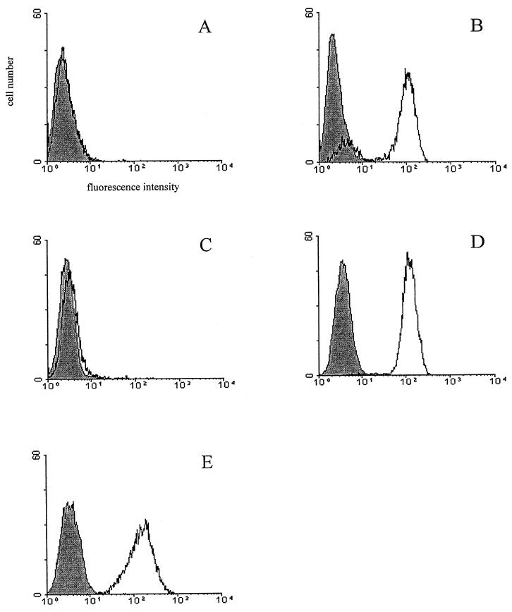
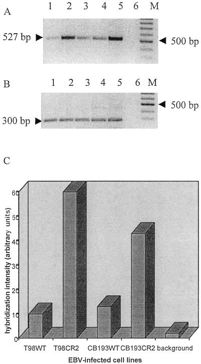
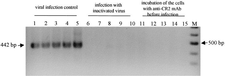
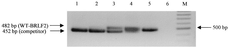
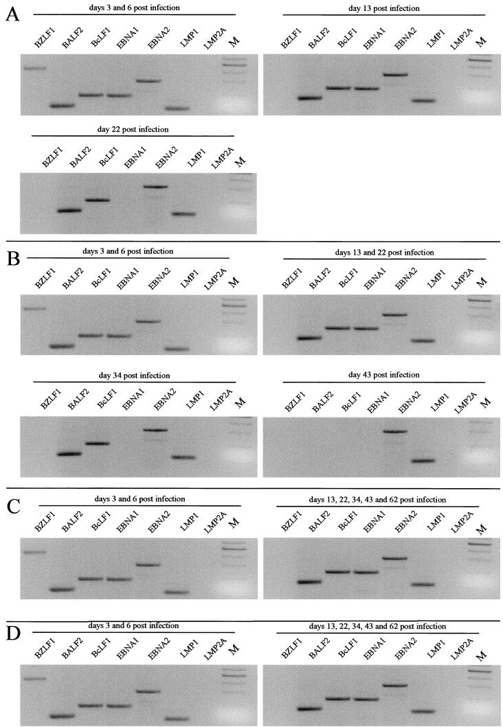
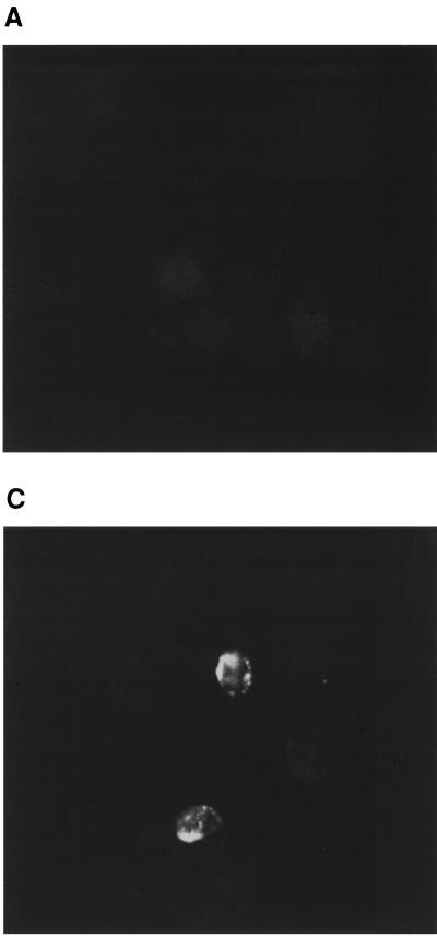
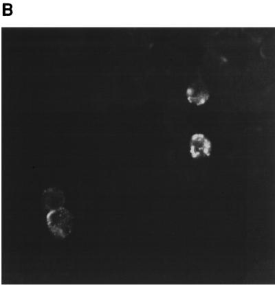
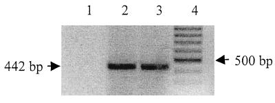
Epstein-Barr virus (EBV) is implicated in different central nervous system syndromes. The major cellular receptor for EBV, complement receptor type 2 (CR2) (CD21), is expressed by different astrocyte cell lines and human fetal astrocytes, suggesting their susceptibility to EBV infection. We demonstrated the infection of two astrocyte cell lines, T98 and CB193, at low levels. As infection was mediated by CR2, we used two stable CR2 transfectant astrocyte cell lines (T98CR2 and CB193CR2) to achieve a more efficient infection. We have monitored EBV gene expression for 2 months and observed the transient infection of T98 and T98CR2 cells and persistent infection of CB193 and CB193CR2 cells. The detection of BZLF1, BALF2, and BcLF1 mRNA expression suggests that the lytic cycle is initiated at early time points postinfection. At later time points the pattern of mRNA expressed (EBER1, EBNA1, EBNA2, and LMP1) differs from latency type III in the absence of LMP2A transcription and in the expression of BALF2 and BcLF1 but not BZLF1. A reactivation of the lytic cycle was achieved in CB193CR2 cells by the addition of phorbol esters. These studies identify astrocyte cell lines as targets for EBV infection and suggest that this infection might play a role in the pathology of EBV in the brain.


Figures

FIG. 1
FIG. 1
Analysis of CR2 expression by using MAb HB5 on wild-type and CR2-transfected astrocyte lines: T98WT (A), T98CR2 (B), CB193WT (C), CB193CR2 (D), and Raji (positive control) (E). The cells were stained with anti-CR2 MAb HB5 (open area) or with an isotype control (shaded area) and FITC-labeled goat F(ab′)2 anti-mouse IgG, as described in Materials and Methods.
FIG. 2
FIG. 2
Detection of EBV DNA on astrocyte lines 48 h after infection. DNA from each cell line was prepared and used as a template for PCR amplification of a 527-bp fragment of the BamHI W fragment of the EBV genome. (A) PCR amplification products were resolved on a 2% agarose gel and detected by staining with ethidium bromide (inverted picture). A negative control that lacked template DNA (lane 6) and a positive control that used B95-8 DNA as a template (lane 5) were included. Lane 1, T98WT; lane 2, T98CR2; lane 3, CB193WT; lane 4, CB193CR2; M, molecular weight marker (100-bp ladder). (B) The same DNA preparation was amplified for the GAPDH gene by using specific primers. (C) Semiquantification of the EBV DNA PCR signal. After Southern blotting, the membrane was hybridized with a specific 32P-labeled probe. The hybridization signal was analyzed with a phosphorimager and calculated as arbitrary units. The background lane shows the hybridization signal from the negative control. The signal obtained for the positive control was too strong, and the quantification was impossible.
FIG. 3
FIG. 3
Expression of spliced BZLF1 mRNA 2 days after infection. Cells were infected with EBV prepared from B95-8 supernatant and harvested 2 days p.i. RNAs were extracted and submitted to PCR amplification with specific primers for BZLF1 mRNAs (their sequences are shown in Table 1). PCR amplification products were detected by staining with ethidium bromide (inverted picture). Different virus preparations were used: EBV concentrated from B95-8 supernatant and diluted in Ham’s F-12 medium supplemented with 2% FCS (lane 1, T98WT; lane 2, T98CR2; lane 3, CB193WT; lane 4, CB193CR2; the B-cell line Ramos was used as a positive control for the infection procedure in lane 5), virus inactivated by boiling for 45 min (lane 6, T98WT; lane 7, T98CR2; lane 8, CB193WT; lane 9, CB193CR2; lane 10, Ramos), or cells preincubated 1 h at 4°C with anti-CR2 MAb FE8 (20 μg/ml) before infection with active viral preparation (lane 11, T98WT; lane 12, T98CR2; lane 13, CB193WT; lane 14, CB193CR2; lane 15, Ramos). M, molecular weight marker (100-bp ladder).
FIG. 4
FIG. 4
Competitive DNA PCR for quantification of viral particles in EBV preparations used for infection. DNA was extracted from the EBV preparation and used for BLRF2 PCR. For quantification a competitor DNA with a 30-bp deletion was added in 10-fold dilutions ranging from 15 × 105 to 15 × 101 molecules per reaction (lanes 1 to 5). When competitor and viral DNAs were present in similar concentrations (here approximately 15 × 103 [lane 3]) both PCR products were visible in similar amounts. Lane 6 shows the PCR negative control. M, molecular weight marker (100-bp ladder). WT, wild type.
FIG. 5
FIG. 5
PCR analysis of BALF2 and BcLF1 mRNAs and spliced BZLF1, EBNA1, EBNA2, LMP1, and LMP2A mRNAs in T98WT (A), T98CR2 (B), CB193WT (C), and CB193CR2 (D) cell lines at different time points after infection. After nested PCR with specific primers the PCR products were detected by staining with ethidium bromide (inverted picture). The sizes expected are 442 bp for BZLF1, 117 bp for BALF2, 181 bp for BcLF1, 172 bp for EBNA1, 300 bp for EBNA2, and 280 bp for LMP2A (see Materials and Methods for details). M, 100-bp ladder with the prominent band of 500 bp.
FIG. 6
FIG. 6
Detection of LMP1 by immunofluorescence. EBV-infected CB193CR2 cells were fixed in 4% paraformaldehyde for 20 min and incubated for 60 min at room temperature with MAb CS1-4, followed by goat anti-mouse IgG. (A) LMP1 staining of uninfected cells. (B) Staining of cells after infection with EBV prepared from B95-8 cell line. (C) Staining of cells 20 days after infection with γ-irradiated B95-8 cells (magnification, ×63).
FIG. 6
FIG. 6
Detection of LMP1 by immunofluorescence. EBV-infected CB193CR2 cells were fixed in 4% paraformaldehyde for 20 min and incubated for 60 min at room temperature with MAb CS1-4, followed by goat anti-mouse IgG. (A) LMP1 staining of uninfected cells. (B) Staining of cells after infection with EBV prepared from B95-8 cell line. (C) Staining of cells 20 days after infection with γ-irradiated B95-8 cells (magnification, ×63).
FIG. 7
FIG. 7
Expression of BZLF1 mRNA 3 and 6 days after stimulation of CB193CR2 cell line (infected with EBV 70 days before) with TPA (5 × 10⁻⁸ M final concentration). RNA was extracted and submitted to PCR amplification with specific primers for BZLF1 mRNAs (the sequences are shown in Table 1). PCR amplification products were detected by staining with ethidium bromide (inverted picture). RT-PCR results from EBV-infected CB193CR2 cells without stimulation (lane 1), after 3 days of stimulation (lane 2), or after 6 days of stimulation (lane 3) are shown. M, molecular weight marker (100-bp ladder).

Similar articles

Cited by

References

    1. Ahearn J M, Fearon D T. Structure and function of the complement receptors, CR1 (CD35) and CR2 (CD21) Adv Immunol. 1989;46:183–219. - PubMed
    1. Alfieri C, Birkenbach M, Kieff E. Early events in Epstein-Barr virus infection of human B lymphocytes. Virology. 1991;181:595–608. - PubMed
    1. Bagasra O, Lavi E, Bobroski L, Khalili K, Pestaner J P, Tawadros R, Pomerantz R J. Cellular reservoirs of HIV-1 in the central nervous system of infected individuals: identification by the combination of in situ polymerase chain reaction and immunohistochemistry. AIDS. 1996;10:573–585. - PubMed
    1. Bogedain C, Alliger P, Schwarzmann F, Marschall M, Wolf H, Jilg W. Different activation of Epstein-Barr virus immediate-early and early genes in Burkitt lymphoma cells and lymphoblastoid cell lines. J Virol. 1994;68:1200–1203. - PMC - PubMed
    1. Bray P F, Culp K W, McFarlin D E, Panitch H S, Torkelson R D, Schlight J P. Demyelinating disease after neurologically complicated primary Epstein-Barr virus infection. Neurology. 1992;42:278–282. - PubMed

Publication types

MeSH terms