. 1999 Sep;73(9):7769-79.
doi: 10.1128/JVI.73.9.7769-7779.1999.

Nuclear export factor CRM1 interacts with nonstructural proteins NS2 from parvovirus minute virus of mice

Affiliations

Nuclear export factor CRM1 interacts with nonstructural proteins NS2 from parvovirus minute virus of mice

U Bodendorf et al. J Virol. 1999 Sep.

Abstract

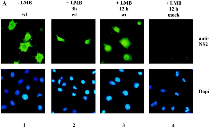
The nonstructural NS2 proteins of autonomous parvoviruses are known to act in a host cell-dependent manner and to play a role in viral DNA replication, efficient translation of viral mRNA, and/or encapsidation. Their exact function during the parvovirus life cycle remains, however, still obscure. We report here the characterization of the interaction with the NS2 proteins from the parvovirus minute virus of mice (MVM) and rat as well as mouse homologues of the human CRM1 protein, a member of the importin-beta family recently identified as an essential nuclear export factor. Using the two-hybrid system, we could detect the interaction between the carboxy-terminal region of rat CRM1 and each of the three isoforms of NS2 (P [or major], Y [or minor], and L [or rare]). NS2 proteins were further shown to interact with the full-length CRM1 by coimmunoprecipitation experiments using extracts from both mouse and rat cell lines. Our data show that CRM1 preferentially binds to the nonphosphorylated isoforms of NS2. Moreover, we observed that the treatment of MVM-infected cells with leptomycin B, a drug that specifically inhibits the CRM1-dependent nuclear export pathway, leads to a drastic accumulation of NS2 proteins in the nucleus. Both NS2 interaction with CRM1 and nuclear accumulation upon leptomycin B treatment strongly suggest that these nonstructural viral proteins are actively exported out of the nuclei of infected cells via a CRM1-mediated nuclear export pathway.


Figures

FIG. 1
FIG. 1
All three NS2 isoforms interact with the C-terminal part of rat CRM1 in the two-hybrid system. The specific interaction of MVM NS2P (A and B), NS2Y (B), or NS2L (B) isoform with the C-terminal domain of the rat crm1 gene product was revealed by means of β-galactosidase assays. SFY526 yeast cells expressing the indicated bait and prey pairs were streaked onto Trp Leu selective dishes containing X-Gal and grown for 48 h at 30°C. A blue or a white color indicates a positive or negative interaction, respectively.
FIG. 2
FIG. 2
Nucleotide sequence of the cDNA encoding the C-terminal domain of the rat crm1 gene product and, below it, the deduced amino acid sequence given in single-letter code. Nucleotides in italics (numbered −15 to −1) indicate the end of the sequence encoding the Gal4-activating domain, at its site of fusion with the isolated cDNA. The polyadenylation site present at the end of the cDNA sequence is underlined. This sequence has received the GenBank accession no. AJ238278.
FIG. 3
FIG. 3
Carboxy-terminal domains of CRM1 proteins are conserved throughout evolution. The alignment of the translated rat CRM1 C-terminal sequence (r) with those from humans (h), fission yeast (f), and budding yeast (b) is shown. White letters represent identical amino acids between the rat sequence and the sequence of at least one other species.
FIG. 4
FIG. 4
Specific coimmunoprecipitation of NS2 and full-length CRM1 proteins from MVM-infected FREJ4 cell extracts. Lysates of 35S-labeled FREJ4 cells, infected (i.) or not infected (n.i.) with MVM at an MOI of 10 PFU per cell, were prepared in the presence (+) or absence (−) of SDS and immunoprecipitated with antisera directed against either the NS2 C terminus (SP6) (A and B), the NS1 C terminus (SP8) (A), or the hCRM1 C terminus (anti-hCRM1) (B). The autoradiograms show immunoprecipitated materials after separation by electrophoresis through 10% (A) or bipartite 6 to 10% (B) SDS-polyacrylamide gels. Arrows to the left point at phosphorylated (p) and nonphosphorylated (np) NS2 (25 kDa), 14-3-3 (30 and 32 kDa), NS1 (83 kDa), and CRM1 (110 kDa) proteins. M, molecular sizes (in kilodaltons) of prestained protein standards.
FIG. 4
FIG. 4
Specific coimmunoprecipitation of NS2 and full-length CRM1 proteins from MVM-infected FREJ4 cell extracts. Lysates of 35S-labeled FREJ4 cells, infected (i.) or not infected (n.i.) with MVM at an MOI of 10 PFU per cell, were prepared in the presence (+) or absence (−) of SDS and immunoprecipitated with antisera directed against either the NS2 C terminus (SP6) (A and B), the NS1 C terminus (SP8) (A), or the hCRM1 C terminus (anti-hCRM1) (B). The autoradiograms show immunoprecipitated materials after separation by electrophoresis through 10% (A) or bipartite 6 to 10% (B) SDS-polyacrylamide gels. Arrows to the left point at phosphorylated (p) and nonphosphorylated (np) NS2 (25 kDa), 14-3-3 (30 and 32 kDa), NS1 (83 kDa), and CRM1 (110 kDa) proteins. M, molecular sizes (in kilodaltons) of prestained protein standards.
FIG. 5
FIG. 5
Association of CRM1 with the nonphosphorylated forms of NS2 in MVM-infected mouse cell extracts. 35S-labeled extracts were prepared from A9 cells that were mock treated (n.i.) or infected (i.) with either wild-type (wt) MVM (A and B) or mutant (NS2-T149A) MVM (B). Immunoprecipitation reactions were performed with SP6 antiserum (A and B) or anti-hCRM1 antibodies (A) in the presence (+) or absence (−) of SDS and analyzed as described in the legend to Fig. 4. Upper and lower panels in panel B correspond to autoradiograms of the same gel exposed for long and short times, respectively. Arrows to the left point at phosphorylated (p) and nonphosphorylated (np) NS2, 14-3-3, and CRM1 proteins. M, molecular sizes (in kilodaltons) of prestained protein standards.
FIG. 6
FIG. 6
Subcellular distribution of NS2 in MVM-infected mouse cells. A9 cells blocked at the G1-to-S transition were infected with MVM and released into S phase by incubation for either 10 or 12 h in a complete medium containing (+ LMB) or not containing (− LMB) 10 nM leptomycin B (LMB). The drug was given either for the whole duration of release (12 h) or during the last 3 h of a 10-h release period (3 h). Cells were further analyzed by indirect immunofluorescence assay with the NS2-specific SP6 serum (A) or the NS1-specific 3D9 antibody (B). Magnification, ×270. wt, wild type.
FIG. 6
FIG. 6
Subcellular distribution of NS2 in MVM-infected mouse cells. A9 cells blocked at the G1-to-S transition were infected with MVM and released into S phase by incubation for either 10 or 12 h in a complete medium containing (+ LMB) or not containing (− LMB) 10 nM leptomycin B (LMB). The drug was given either for the whole duration of release (12 h) or during the last 3 h of a 10-h release period (3 h). Cells were further analyzed by indirect immunofluorescence assay with the NS2-specific SP6 serum (A) or the NS1-specific 3D9 antibody (B). Magnification, ×270. wt, wild type.

References

    1. Adachi Y, Yanagida M. Higher order chromosome structure is affected by cold-sensitive mutations in a Schizosaccharomyces pombe gene, crm1+, which encodes a 115-kDa protein preferentially localized to the nucleus and its periphery. J Cell Biol. 1989;108:1195–1207. - PMC - PubMed
    1. Anouja F, Wattiez R, Mousset S, Caillet-Fauquet P. The cytotoxicity of the parvovirus minute virus of mice nonstructural protein NS1 is related to changes in the synthesis and phosphorylation of cell proteins. J Virol. 1997;71:4671–4678. - PMC - PubMed
    1. Askjaer P, Jensen T H, Nilsson J, Englmeier L, Kjems J. The specificity of the CRM1-Rev nuclear export signal interaction is mediated by RanGTP. J Biol Chem. 1998;273:33414–33422. - PubMed
    1. Ballaun C, Farrington G K, Dobrovnik M, Rusche J, Hauber J, Böhnlein E. Functional analysis of human T-cell leukemia virus type 1 Rex response element: direct RNA binding of Rex protein correlates with in vivo activity. J Virol. 1991;65:4408–4413. - PMC - PubMed
    1. Bartel P L, Chien C-T, Sternglanz R, Fields S. Using the two-hybrid system to detect protein-protein interactions. In: Hartley D A, editor. Cellular interactions in development: a practical approach. Oxford, United Kingdom: Oxford University Press; 1993. pp. 153–179.

Publication types

MeSH terms

Associated data