

doi: 10.1128/JVI.73.9.7874-7876.1999.
Rhesus rhadinovirus establishes a latent infection in B lymphocytes in vivo
Affiliations
- PMID: 10438883
- PMCID: PMC104320
- DOI: 10.1128/JVI.73.9.7874-7876.1999
Item in Clipboard
Rhesus rhadinovirus establishes a latent infection in B lymphocytes in vivo
J Virol.
1999 Sep.
Abstract
Recent DNA sequence analysis indicates that rhesus rhadinovirus (RRV) is a member of the lymphotropic gamma-2 herpesvirus family. To determine if RRV is lymphotropic, peripheral blood mononuclear cells from naturally infected monkeys were separated by immunomagnetic bead depletion and analyzed for the presence of RRV by virus isolation and nested PCR. The recovery and consistent detection of RRV in the CD20(+)-enriched fraction clearly demonstrates that B lymphocytes are a major site of virus persistence.
Figures
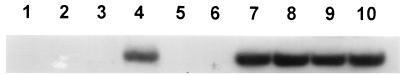
RRV DNA detection by nested PCR amplification of PBMCs obtained from healthy rhesus monkeys. Total DNA from purified PBMCs was amplified by nested PCR, and the products were analyzed by Southern hybridization. Lanes 1 to 3, PCR reagent controls containing water, 50 pg of rhesus cytomegalovirus DNA, or 100 ng of rhesus lymphocryptovirus-infected cell DNA, respectively; lanes 4 to 10, reaction mixtures containing PBMC DNA from healthy seropositive monkeys (lanes 4, 7, 8, 9, and 10) or seronegative monkeys (lanes 5 and 6).
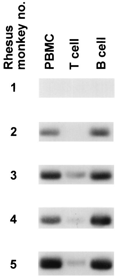
Detection of RRV DNA in enriched lymphocyte fractions. Total DNA from PBMCs, T-cell-enriched (T cell) or B-cell-enriched (B cell) fractions, obtained from the monkeys indicated in Table 2, were amplified by nested PCR, and the products were analyzed by Southern hybridization.
References
-
- Ambroziak J A, Blackbourn D J, Herndier B G, Glogau R G, Gullett J H, McDonald A R, Lennette E T, Levy J A. Herpes-like sequences in HIV-infected and uninfected Kaposi’s sarcoma patients. Science. 1995;268:582–583. - PubMed
-
- Boshoff C, Schulz T F, Kennedy M M, Graham A K, Fisher C, Thomas A, McGee J O, Weiss R A, O’Leary J J. Kaposi’s sarcoma-associated herpesvirus infects endothelial and spindle cells. Nat Med. 1995;1:1274–1278. - PubMed
-
- Cesarman E, Chang Y, Moore P S, Said J W, Knowles D M. Kaposi’s sarcoma-associated herpesvirus-like DNA sequences in AIDS-related body cavity-based lymphomas. N Engl J Med. 1995;332:1186–1191. - PubMed
-
- Chang Y, Cesarman E, Pessin M S, Lee F, Culpepper J, Knowles D M, Moore P S. Identification of herpesvirus-like DNA sequences in AIDS-associated Kaposi’s sarcoma. Science. 1994;266:1865–1869. - PubMed
Publication types
MeSH terms
Substances
Grants and funding
LinkOut - more resources
Full Text Sources
