

doi: 10.1128/JVI.73.9.7882-7885.1999.
Middle T antigen activation of signal transduction pathways does not overcome p53-mediated growth arrest
Affiliations
- PMID: 10438885
- PMCID: PMC104322
- DOI: 10.1128/JVI.73.9.7882-7885.1999
Item in Clipboard
Middle T antigen activation of signal transduction pathways does not overcome p53-mediated growth arrest
J Virol.
1999 Sep.
Abstract
Polyomavirus middle T antigen does not overcome p53-mediated G(1) arrest in mouse embryo fibroblasts. Middle T antigen still associates with the signaling molecules phosphatidylinositol 3-kinase and SHC and activates the transcriptional activity of c-Myc and AP1 in p53-arrested cells. Examination of cell cycle regulatory proteins indicated that p53 does not interfere with these mitogenic signals but acts later in the G(1) phase of the cell cycle.
Figures
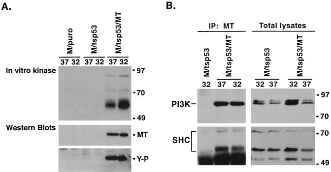
MT associates with a tyrosine kinase and interacts with PI3K and SHC in p53 growth-arrested cells. (A) Extracts were prepared from M/puro, M/tsp53, and M/tsp53/MT cells cultured at 37 and 32°C. MT was immunoprecipitated and incubated with [γ-32P]ATP to detect associated kinase activity. The autoradiographs displayed in the upper panel show the MT-associated kinase from designated cells cultured at indicated temperatures (°C). The lower panel shows Western blots of total-cell lysates probed with antibodies to MT and antibodies to phosphotyrosine (Y-P). (B) Extracts were prepared from M/tsp53 and M/tsp53/MT cells cultured at 37 or 32°C. Immunoblots of MT immune complexes (IP MT) were probed with antibodies to PI3K or SHC (left panels). As a control, Western blots of total-cell lysate were probed to detect cellular levels of PI3K and SHC at each temperature (°C) (right panels). Numbers at right of each panel indicate molecular masses in kilodaltons.
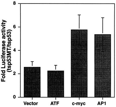
Transcriptional activity driven by c-Myc and AP1 is greater in p53 growth-arrested cells expressing MT. M/tsp53 and M/tsp53/MT cells were transfected with luciferase reporter vectors containing multiple ATF, c-myc, or AP1 binding sites upstream of a minimal promoter. The luciferase reporter plasmid with the minimal promoter was also transfected (Vector). A reporter plasmid containing the rat β-actin promoter driving expression of β-galactosidase was cotransfected and used as a control for transfection efficiency. Following transfection, cells were cultured in Dulbecco modified Eagle medium plus 0.5% serum at 37°C for 24 h and then at 32°C for 48 h. Cell extracts were harvested, and luciferase and β-galactosidase activities were measured. The graph compares luciferase activity in M/tsp53/MT cells to that in M/tsp53 cells. Each transfection was done in duplicate. The data represent the averages of four independent experiments. The error bars represent standard deviations between experiments.
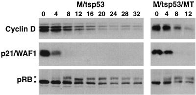
p53 arrests cells in late G1 phase, after expression of cyclin D. M/tsp53 and M/tsp53/MT cells were grown at 32°C for 24 h to arrest growth (0), followed by culture at 37°C for the times (hours) indicated above the panels. Cell extract was made, and protein levels were analyzed by Western blotting. Blots were probed with antibodies to cyclin D, p21/WAF1, and pRB.
References
-
- Bates S, Vousden K H. p53 in signaling checkpoint arrest and apoptosis. Curr Opin Genet Dev. 1996;6:12–19. - PubMed
-
- Benjamin T, Vogt P K. Cell transformation by viruses. In: Fields B, editor. Virology. New York, N.Y: Raven Press; 1990. pp. 345–367.
-
- Deng C, Zhang P, Harper J W, Elledge S J, Leder P. Mice lacking p21/CIP1/WAF1 undergo normal development, but are defective in G1 checkpoint control. Cell. 1995;82:675–684. - PubMed
-
- Dilworth S M, Brewster C E P, Jones M D, Lanfrancone L, Pelicci G, Pelicci P G. Transformation by polyoma virus middle T-antigen involves the binding and tyrosine phosphorylation of Shc. Nature. 1994;367:87–90. - PubMed
Publication types
MeSH terms
Substances
Grants and funding
LinkOut - more resources
Full Text Sources
Research Materials
Miscellaneous
