. 1999 Aug 9;146(3):559-72.
doi: 10.1083/jcb.146.3.559.

Localization in the nucleolus and coiled bodies of protein subunits of the ribonucleoprotein ribonuclease P

Affiliations

Localization in the nucleolus and coiled bodies of protein subunits of the ribonucleoprotein ribonuclease P

N Jarrous et al. J Cell Biol. .

Abstract

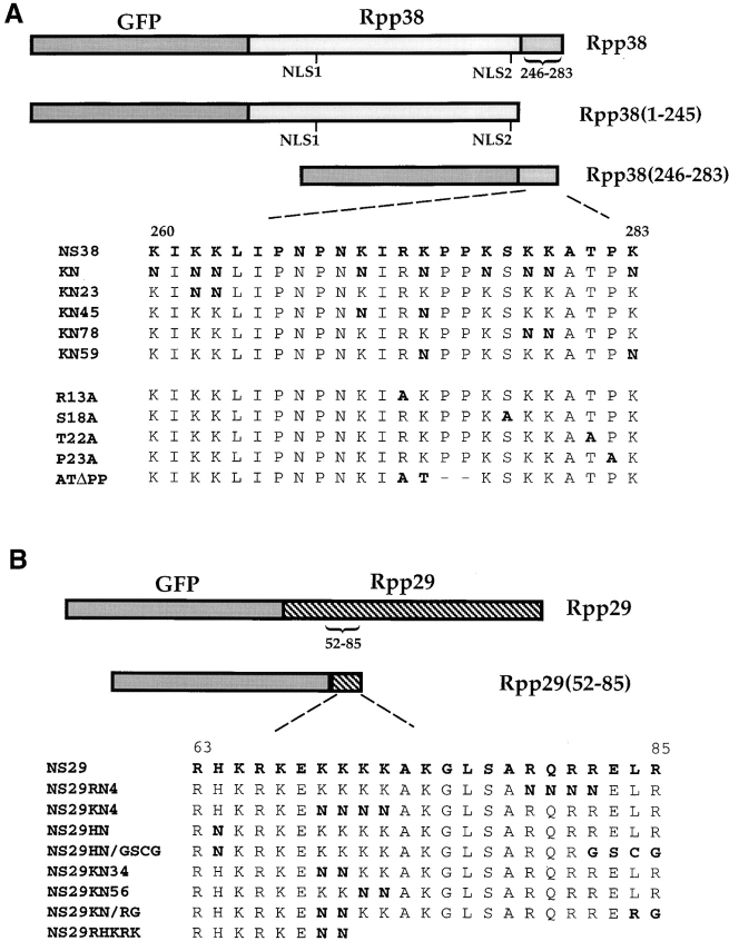
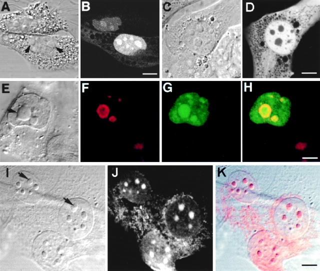
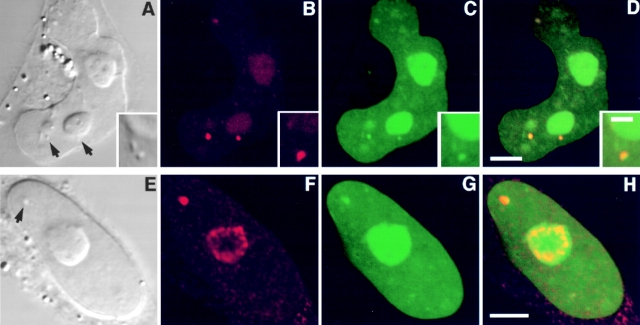
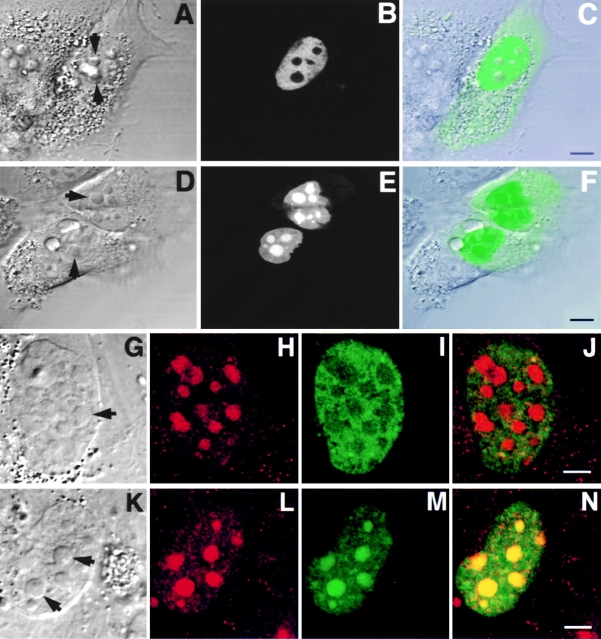
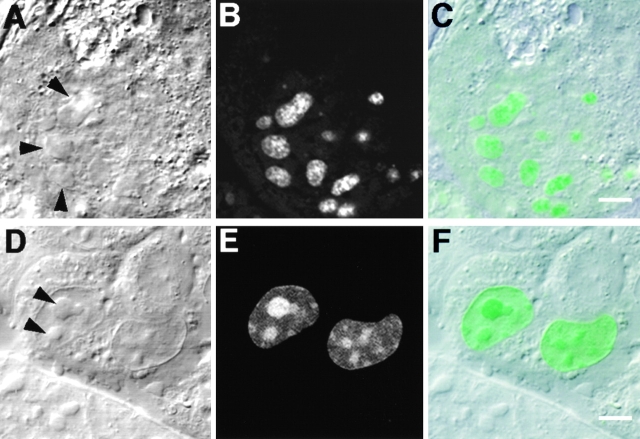
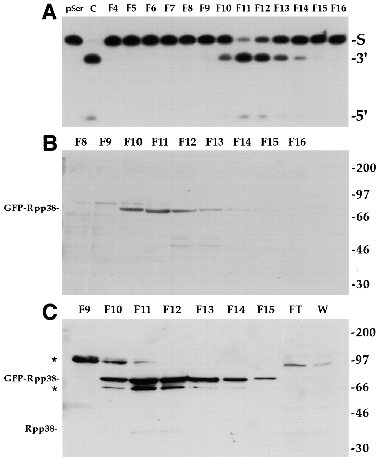
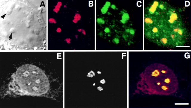
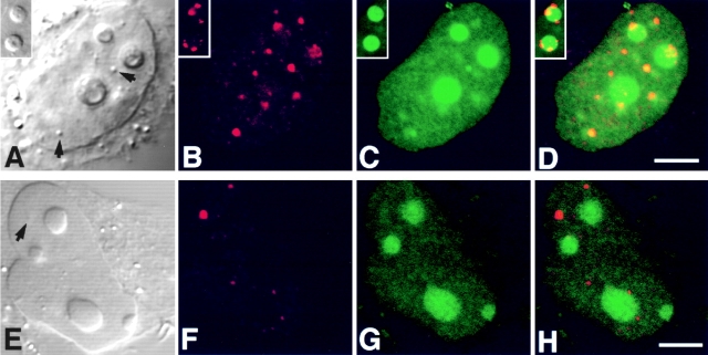
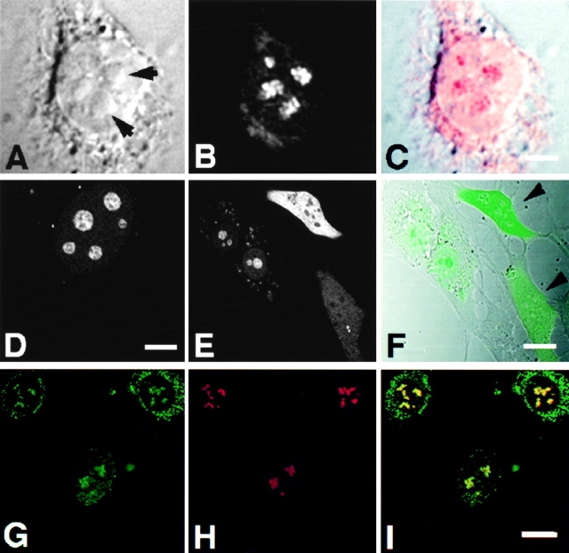
The precise location of the tRNA processing ribonucleoprotein ribonuclease P (RNase P) and the mechanism of its intranuclear distribution have not been completely delineated. We show that three protein subunits of human RNase P (Rpp), Rpp14, Rpp29 and Rpp38, are found in the nucleolus and that each can localize a reporter protein to nucleoli of cells in tissue culture. In contrast to Rpp38, which is uniformly distributed in nucleoli, Rpp14 and Rpp29 are confined to the dense fibrillar component. Rpp29 and Rpp38 possess functional, yet distinct domains required for subnucleolar localization. The subunit Rpp14 lacks such a domain and appears to be dependent on a piggyback process to reach the nucleolus. Biochemical analysis suggests that catalytically active RNase P exists in the nucleolus. We also provide evidence that Rpp29 and Rpp38 reside in coiled bodies, organelles that are implicated in the biogenesis of several other small nuclear ribonucleoproteins required for processing of precursor mRNA. Because some protein subunits of RNase P are shared by the ribosomal RNA processing ribonucleoprotein RNase MRP, these two evolutionary related holoenzymes may share common intranuclear localization and assembly pathways to coordinate the processing of tRNA and rRNA precursors.


Figures

Figure 1
Figure 1
Schematic representation of gene constructs. (A) Rpp38 or portions of this protein, Rpp38(1-245) or Rpp38(246-283), was fused in-frame to GFP in pEGFP-C1 expression vector (see Materials and Methods). Numbers indicate positions of residues in the 283–amino acid Rpp38 polypeptide. NLS1 and NLS2 indicate putative NLSs. NS38 represents the amino acid sequence from position 260–283 of Rpp38. Substitution mutations of the nine lysines to asparagines in NS38 sequence, and their numbers, are presented in bold letters. Single substitution mutants of arginine (R13A), serine (S18A), threonine (T22A), and proline (P23A) to alanine are also shown. ATΔPP has substitution of arginine and lysine with alanine and threonine, respectively, and the two consecutive proline residues were deleted from NS38. (B) Full-length Rpp29 or the amino acids encompassing positions 52–85 or 63–85 of Rpp29 were fused in-frame with GFP in pEGFP-C1. The amino acid sequence of Rpp29 from position 63–85, designated NS29, is indicated. Several mutants of NS29 with single and multiple substitutions or deletions are shown. All gene fusion constructs were transcribed from a cytomegalovirus promoter-enhancer located upstream of GFP in the expression plasmid.
Figure 2
Figure 2
Intracellular localization of Rpp38. Swiss 3T3 fibroblasts were transiently transfected (see Materials and Methods) with pEGFP-Rpp38 (A and B) or pEGFP-C1 (C and D). 48 h after transfection, living cells were examined with a confocal microscope. Colocalization of GFP-Rpp38 in the nucleolus with the protein B23 in transfected HeLa cells is demonstrated by indirect immunofluorescence analysis (E–H). (I–K) Immunofluorescence of endogenous Rpp38 protein in untransfected 3T3 fibroblasts using anti-Rpp38 antibodies showing uniformly stained nucleoli. Images of DIC (A, C, E, and I), GFP (B, D, and G), B23 (F), and anti-Rpp38 (J) are shown. H is an overlay of F and G, and K is an overlay of I and J. No specific signal above autofluorescence was seen when control sera of rabbits were tested (not shown). Arrows point to nucleoli. Bars: B, D, and K, 5 μm; H, 2.5 μm.
Figure 3
Figure 3
GFP-Rpp38 is localized in the nucleoli and coiled bodies. HeLa cells were transfected for 48 h with pEGFP-Rpp38, and then were examined for the presence of p80-coilin (A–D) or Nopp140 (E–H) in indirect immunofluorescence analysis. DIC (A and E), p80-coilin (B, red), Nopp140 (F, red), GFP-Rpp38 (C and G, green), and the overlays of B over C and F over G are shown in D and H, respectively. Intense yellow color is seen in the nucleoli and coiled bodies indicated by arrows. Overlays were acquired at the same confocal plane using Texas red (red) and FITC (green) filters. Bleedthrough between the two channels was completely eliminated. 3–6 coiled bodies were usually seen in HeLa cells. Inserts shown in A–D represent higher magnification of coilin-immunostained coiled bodies in proximity to a nucleolus of HeLa cells expressing GFP-Rpp38. All images were acquired during the same experimental observation. Bars: D and H, 2 μm; insert, 0.5 μm.
Figure 4
Figure 4
A nucleolar localization signal exists in the carboxy terminus of Rpp38. Swiss 3T3 fibroblasts were transfected with pEGFP-Rpp38(1-245) (A–C and G–J) or with pEGFP-Rpp38(246-283) (D–F and K–N). Construct maps are shown in Fig. 1 A. 48 h after transfection, living cells were examined (A–F) using confocal microscopy, or first fixed and subjected to indirect immunofluorescence using antibody against nucleolar protein B23 (G–J and K–N). DIC (A, D, G, and K), GFP (B, E, I, and M, green), B23 (H and L, red), and overlays (C, F, J, and N) are shown. Arrows point to nucleoli. Bars: C and F, 5 μm; J and N, 2.5 μm.
Figure 6
Figure 6
Subnucleolar localization of GFP-Rpp29 and identification of NS29. Swiss 3T3 fibroblasts were transiently transfected with pEGFP-Rpp29 (A–C) or pEGFP-Rpp29(52-85) (D–F). 48 h after transfection, localization of the fusion proteins was determined by confocal microscopy. DIC (left) GFP (middle), and overlay (right) images are shown. Arrows indicate nucleoli. Bars: C and F, 3.3 and 5 μm, respectively.
Figure 5
Figure 5
GFP-Rpp38 is associated with active RNase P. (A) S100 crude extracts obtained from of G418-resistant, stably transfected 293 HEK cell populations were fractionated on a DEAE-Sepharose anion exchange column (see Materials and Methods) and fractions eluted from the column were tested for RNase P activity using the yeast suppressor precursor tRNASer (S) as substrate (left lane). RNase P obtained from untransfected cells was used as a positive control (second lane from left). The cleavage products, 5′ leader sequence (5′) and mature tRNA (3′), were resolved in 8% polyacrylamide/7 M urea gel. Fraction numbers are indicated above the panel. Equal volumes from the fractions across the RNase P activity tested in A were separated in 12% SDS-PAGE and subjected to Western blot analysis using polyclonal anti-GFP rabbit antibodies (B) or affinity-purified, polyclonal anti-Rpp38 rabbit antibodies (C). FT and W in C stand for concentrated flowthrough and wash fractions of the DEAE column. Positions of GFP-Rpp38, endogenous Rpp38 protein, and protein size markers are shown. Cross-reaction with unknown proteins of M r ∼65 and ∼95 kD are indicated by asterisks.
Figure 7
Figure 7
Colocalization of Rpp29 with B23 and fibrillarin in the nucleolus. Swiss 3T3 fibroblasts were transfected with pEGFP-Rpp29. 48 h later, cells were fixed and subjected to immunostaining with an antibody against nucleolar protein B23 (A–D). Arrows indicate nucleoli. E and F show double immunofluorescence obtained with anti-Rpp29 and antifibrillarin antibodies, respectively, in untransfected 3T3 fibroblasts. G is an overlay of E and F, acquired at the same confocal plane using Texas red (red) or FITC (green) filters. An intense yellow color is seen in nucleoli. Bleedthrough between the two channels was completely eliminated. Bars: D and G, 2.5 and 5 μm, respectively.
Figure 8
Figure 8
GFP-Rpp29 is localized in nucleoli and coiled bodies. HeLa cells were transfected for 48 h with pEGFP-Rpp29 (A–D) or pEGFP-Rpp14 (E–H), and then immunostained for p80-coilin in indirect immunofluorescence analysis. DIC (A and E), p80-coilin (B and F, red), GFP (C and G, green), and the overlays of B over C and F over G are shown in D and H, respectively. Images in A–D and E–H were acquired at the same confocal plane. The GFP fluorescent signal in C and D was enhanced to highlight the punctate staining seen in the nucleoplasm. Coiled bodies are indicated by arrows. Inserts seen in A–D represent higher magnification of coilin-immunostained coiled bodies in the periphery to two nucleoli of HeLa cells expressing GFP-Rpp29. All images were obtained during the same experimental observation. Bars: D and H, 2.5 μm.
Figure 9
Figure 9
Subnucleolar localization patterns of Rpp14. 3T3 fibroblasts were subjected to indirect immunofluorescent analysis using anti-Rpp14 antibodies (A–C). C is an overlay of A and B. Fibroblasts transfected for 48 h with pEGFP-Rpp14 were examined under confocal microscope (D–F) before fixation and immunofluorescence analysis with anti-Rpp29 antibodies (G–I). F is an overlay of DIC (not shown) and E; two cells with high fluorescent signal are indicated by arrowheads. I is an overlay of G and H. Intense yellow color is seen in nucleoli. Bars: C and D, 3.3 μm; F and I, 10 μm.

References

    1. Andrade L.E.C., Tan E.M., Chan E.K.L. Immunocytochemical analysis of the coiled body in the cell cycle and during cell proliferation. Proc. Natl. Acad. Sci. USA. 1993;90:1947–1951. - PMC - PubMed
    1. Antoine M., Reimers K., Dickson C., Kiefer P. Fibroblast growth factor 3, a protein with dual subcellular localization, is targeted to the nucleus and nucleolus by the concerted action of two nuclear localization signals and a nucleolar retention signal. J. Biol. Chem. 1997;272:29475–29481. - PubMed
    1. Bachant J.B., Elledge S.J. Mitotic treasures in the nucleolus. Nature. 1999;398:757–758. - PubMed
    1. Bauer D.W., Murphy C., Wu Z., Wu C.H., Gall J.G. In vitro assembly of coiled bodies in Xenopus egg extract. Mol. Biol. Cell. 1994;5:633–644. - PMC - PubMed
    1. Bertrand E., Houser-Scott F., Kendall A., Singer R.H., Engelke D.R. Nucleolar localization of early tRNA processing. Genes Dev. 1998;12:2463–2468. - PMC - PubMed

Publication types

MeSH terms