. 1999 Aug 9;146(3):597-608.
doi: 10.1083/jcb.146.3.597.

Cytoplasmic dynein is required for the nuclear attachment and migration of centrosomes during mitosis in Drosophila

Affiliations

Cytoplasmic dynein is required for the nuclear attachment and migration of centrosomes during mitosis in Drosophila

J T Robinson et al. J Cell Biol. .

Abstract

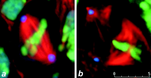
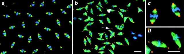
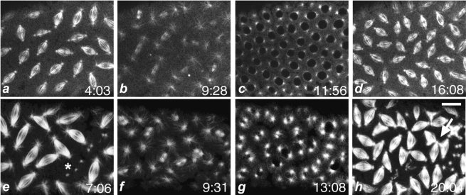
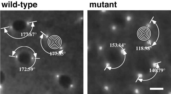
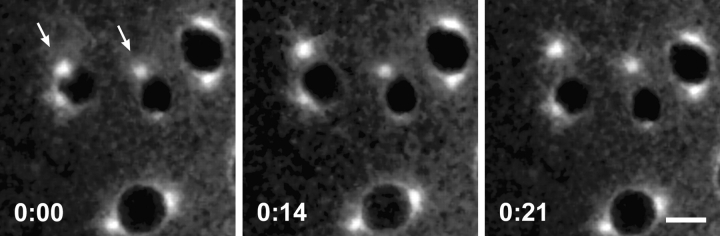
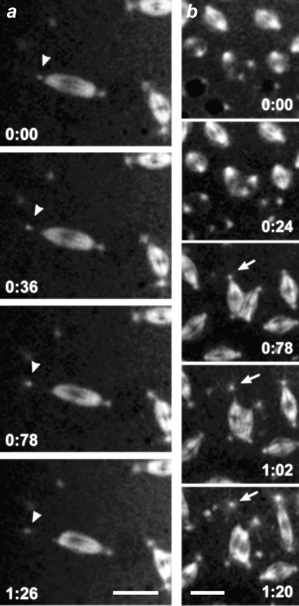
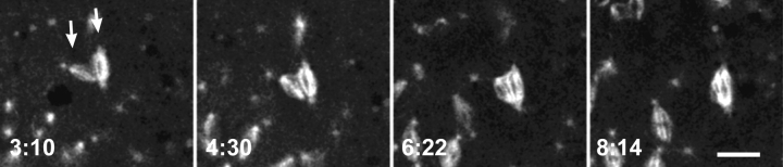
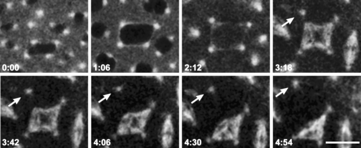
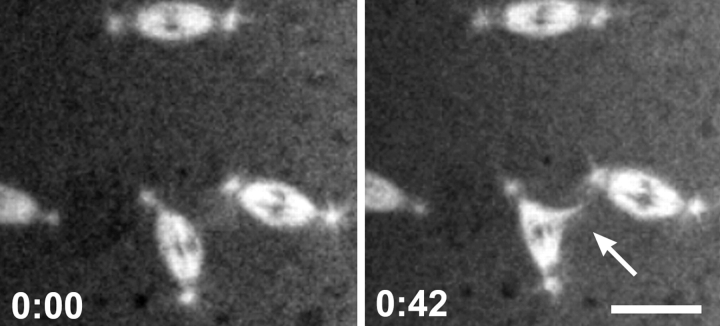
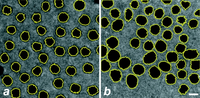
Cytoplasmic dynein is a multisubunit minus-end-directed microtubule motor that serves multiple cellular functions. Genetic studies in Drosophila and mouse have demonstrated that dynein function is essential in metazoan organisms. However, whether the essential function of dynein reflects a mitotic requirement, and what specific mitotic tasks require dynein remains controversial. Drosophila is an excellent genetic system in which to analyze dynein function in mitosis, providing excellent cytology in embryonic and somatic cells. We have used previously characterized recessive lethal mutations in the dynein heavy chain gene, Dhc64C, to reveal the contributions of the dynein motor to mitotic centrosome behavior in the syncytial embryo. Embryos lacking wild-type cytoplasmic dynein heavy chain were analyzed by in vivo analysis of rhodamine-labeled microtubules, as well as by immunofluorescence in situ methods. Comparisons between wild-type and Dhc64C mutant embryos reveal that dynein function is required for the attachment and migration of centrosomes along the nuclear envelope during interphase/prophase, and to maintain the attachment of centrosomes to mitotic spindle poles. The disruption of these centrosome attachments in mutant embryos reveals a critical role for dynein function and centrosome positioning in the spatial organization of the syncytial cytoplasm of the developing embryo.


Figures

Figure 2
Figure 2
Dynein is required for centrosome attachment to spindle poles in larval neuroblasts. Confocal images of Drosophila wild-type and Dhc64C6-10/Df(3L)10H mutant larval neuroblasts taken from a fixed specimen (see Materials and Methods). Similar results were obtained for the genotype Dhc64C6-6/Df(3L)10H. Shown are optical slices of a metaphase neuroblast from wild-type (a) and mutant (b) larval brains. DNA is false-colored in green, α-tubulin is red, and CP190 centrosome antigen is blue. Bar, 5 μm.
Figure 1
Figure 1
Mitotic defects are apparent in fixed mutant dynein Drosophila embryos. Shown are in situ confocal images of wild-type and Dhc64C mutant syncytial embryos. The mutant embryos were collected from Dhc64C6-8/Dhc64C6-6females, fixed, and prepared for immunofluorescence as described (see Materials and Methods). Shown are fields of metaphase nuclei for wild-type (a and c) and Dhc64C mutant (b and d) embryos at similar nuclear division cycles. c and d are enlargements of selected nuclei from a and b, respectively. The arrow points to a free centrosome and the arrowhead indicates a blunt-ended spindle lacking a centrosome. Note that the centrosomes at the periphery of a (e.g., upper left) attach to spindles at the edge of the embryo that tilt out of the image plane. DNA is pseudocolored blue, β-tubulin is green, and γ-tubulin is red. Bars, 10 μm.
Figure 3
Figure 3
Mitotic defects can occur during the early nuclear cycles within dynein mutant embryos. Shown are in situ confocal images of mutant Drosophila syncytial metaphase nuclei. The image was taken from a cycle 3 embryo. The mutant embryos were collected from Dhc64C6-8/Dhc64C6-6females, fixed, and prepared for immunofluorescence as described (see Materials and Methods). a shows a field of mutant metaphase figures with β-tubulin false-colored in green and γ-tubulin in red (overlap appears orange). b shows only the γ-tubulin channel from a.
Figure 4
Figure 4
Dynein mutant embryos display mitotic defects. Time course of Drosophila wild-type and mutant syncytial embryos in vivo by confocal microscopy. The embryos were collected from wild-type Oregon R or Dhc64C6-8/Dhc64C6-6females and microinjected with TRITC-labeled tubulin. Shown are successive confocal images selected from time-lapse collections at the indicated time points (minutes). a–d follow a sequence of mitotic cycles in a representative wild-type embryo, and e–h follow a sequence of mitotic cycles in a typical mutant embryo. Shown is a field of metaphase spindles in a and e, telophase spindles in b and f, prophase spindles in c and g, and the subsequent metaphase in d and h. The arrow points to abnormal spindle interactions. The asterisk marks a patch of free centrosomes. Bar, 10 μm.
Figure 5
Figure 5
Dynein is required for complete centrosome migration. The centrosome migration defect was measured in the manner indicated in both wild-type and dynein mutant embryos (see Materials and Methods). Prophase centrosomes were visualized in vivo by confocal microscopy after microinjection of TRITC-labeled tubulin. The wild-type panel shows a field of syncytial prophase nuclei and associated centrosome separation angles. The mutant panel shows a field of syncytial prophase nuclei and their centrosome separation angles in Dhc64C6-8/Dhc64C6-6embryos. Bar, 5 μm.
Figure 6
Figure 6
Centrosomes can detach from the nuclear envelope in the dynein mutants. Time course of mitosis in a syncytial embryo provided by Dhc64C6-8/Dhc64C6-6females. Microtubules are labeled with TRITC-tubulin. Presented are selected in vivo confocal images that highlight nuclei and labeled centrosomes. The images were taken at the indicated time points (minutes) during prophase. Arrows point to centrosomes, which become separated from the nuclear envelope during the time course shown. Bar, 5 μm.
Figure 7
Figure 7
Centrosomes can detach from metaphase spindles in the dynein mutants. In vivo time course of mitosis during metaphase in a syncytial embryo provided by Dhc64C6-8/Dhc64C6-6females. Shown are selected confocal images of a field containing TRITC-labeled spindles and centrosomes at the indicated time points (minutes). (a) Centrosomes can detach from bipolar spindles. The arrowhead marks a centrosome which detaches and moves away from one pole of a bipolar spindle. (b) Centrosomes can detach from multipolar spindle arrays. The arrow points to a centrosome that detaches and moves away from a multipolar spindle. The associated spindle pole subsequently collapses. Bars, 10 μm.
Figure 8
Figure 8
Fusion of neighboring mitotic arrays in mutant dynein syncytial embryos. Time course of mitosis in vivo within an embryo provided by Dhc64C6-8/Dhc64C6-6females. Microtubules are labeled with TRITC-tubulin. Shown are selected confocal images of spindles at the indicated time points (minutes) during metaphase in vivo within a syncytial embryo. Two spindles (arrows) undergo fusion to form a single bipolar array. Bar, 10 μm.
Figure 9
Figure 9
Alternative pathway for the formation of multipolar spindle arrays. Time-lapse series of confocal images of nuclei in an embryo from Dhc64C6-8/Dhc64C6-6females. Microtubules are labeled with TRITC-labeled tubulin. Shown are syncytial nuclei at the indicated time points (minutes) during the prophase to metaphase transition. A prophase nucleus bearing four centrosomes develops into an abnormal tetrapolar mitotic spindle. A centrosome (arrows) can be seen to detach and move away from one of the poles. This pole subsequently collapses. Bar, 10 μm.
Figure 10
Figure 10
Ectopic spindle pole formation in mutant dynein syncytial embryos. Time points (minutes) of metaphase during mitosis in an embryo provided by Dhc64C6-8/Dhc64C6-6females. Shown are two panels containing metaphase nuclei with spindles visualized with TRITC-tubulin at the time points indicated. The arrow points to an ectopic spindle pole generated between two spindles. The microtubule bundle composing the new pole has split away from the parent spindle. Bar, 10 μm.
Figure 11
Figure 11
Nuclear DNA content is affected in the dynein mutant syncytial nuclei. Shown is an example of the method used (see Materials and Methods) to estimate the relative nuclear volume of both wild-type (a) and dynein mutant (b) Drosophila embryos. Syncytial nuclei are outlined in yellow and false-colored in black. Note variable size and spatial distribution of nuclei in the Dhc mutant embryo. The average diameters of wild-type and dynein mutant nuclei are 11.48 ± 2.58 μm (4 embryos, 428 nuclei), and 13.75 ± 4.47 μm (4 embryos, 437 nuclei), respectively. Bar, 5 μm. The difference in nuclear size is significant (t stat = 3.93; 96% significance).

References

    1. Allan V. Organelle movement. Dynactinportrait of a dynein regulator. Curr. Biol. 1994;4:1000–1002. - PubMed
    1. Boleti H., Karsenti E., Vernos I. Xklp2, a novel Xenopus centrosomal kinesin-like protein required for centrosome separation during mitosis. Cell. 1996;84:49–59. - PubMed
    1. Bruno K.S., Tinsley J.H., Minke P.F., Plamann M. Genetic interactions among cytoplasmic dynein, dynactin, and nuclear distribution mutants of Neurospora crassa . Proc. Natl. Acad. Sci. USA. 1996;93:4775–4780. - PMC - PubMed
    1. Busson S., Dujardin D., Moreau A., Dompierre J., De Mey J.R. Dynein and dynactin are localized to astral microtubules and at cortical sites in mitotic epithelial cells. Curr. Biol. 1998;8:541–544. - PubMed
    1. Callaini G., Dallai R., Riparbelli M.G. Cytochalasin induces spindle fusion in the syncytial blastoderm of the early Drosophila embryo. Biol. Cell. 1992;74:249–254. - PubMed

Publication types

MeSH terms