. 1999 Aug 9;146(3):631-44.
doi: 10.1083/jcb.146.3.631.

I-band titin in cardiac muscle is a three-element molecular spring and is critical for maintaining thin filament structure

Affiliations

I-band titin in cardiac muscle is a three-element molecular spring and is critical for maintaining thin filament structure

W A Linke et al. J Cell Biol. .

Abstract

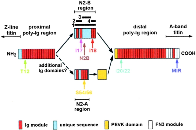
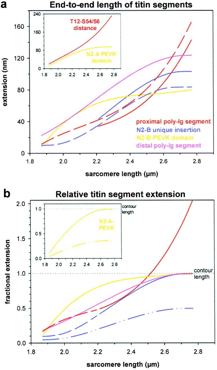
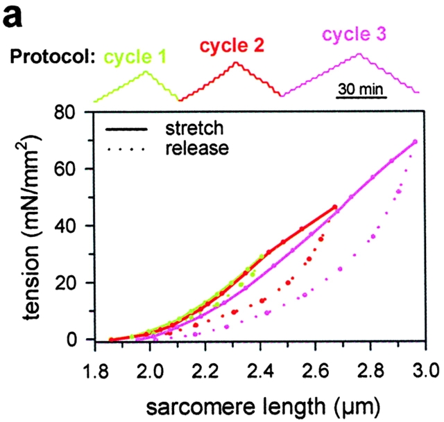
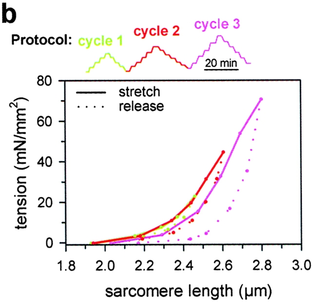
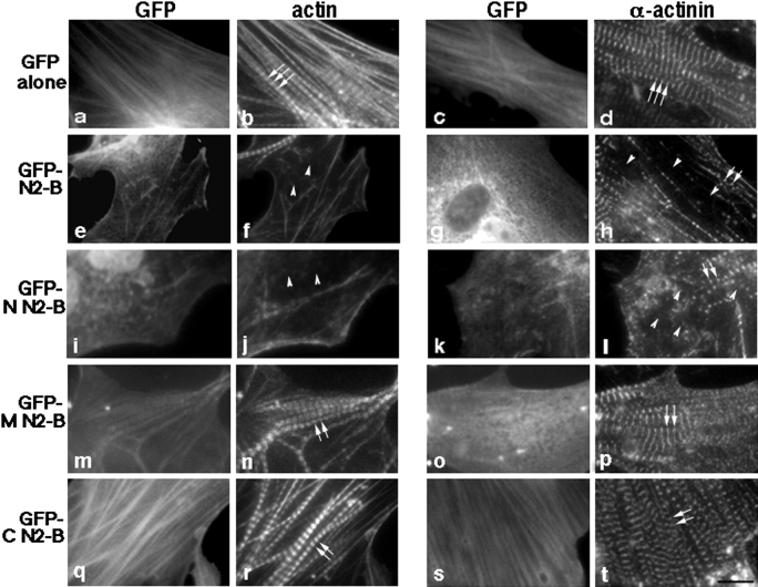
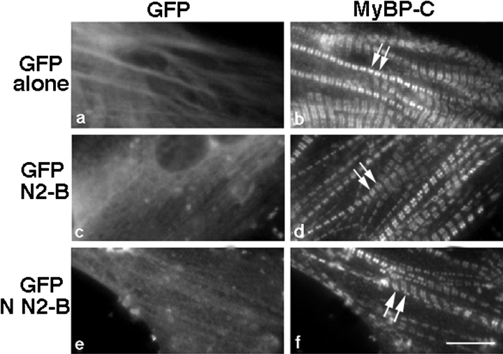
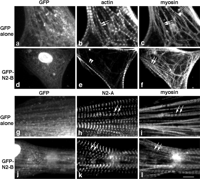
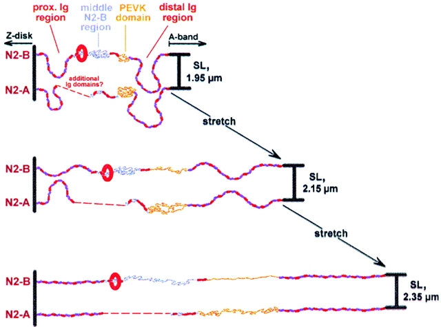
In cardiac muscle, the giant protein titin exists in different length isoforms expressed in the molecule's I-band region. Both isoforms, termed N2-A and N2-B, comprise stretches of Ig-like modules separated by the PEVK domain. Central I-band titin also contains isoform-specific Ig-motifs and nonmodular sequences, notably a longer insertion in N2-B. We investigated the elastic behavior of the I-band isoforms by using single-myofibril mechanics, immunofluorescence microscopy, and immunoelectron microscopy of rabbit cardiac sarcomeres stained with sequence-assigned antibodies. Moreover, we overexpressed constructs from the N2-B region in chick cardiac cells to search for possible structural properties of this cardiac-specific segment. We found that cardiac titin contains three distinct elastic elements: poly-Ig regions, the PEVK domain, and the N2-B sequence insertion, which extends approximately 60 nm at high physiological stretch. Recruitment of all three elements allows cardiac titin to extend fully reversibly at physiological sarcomere lengths, without the need to unfold Ig domains. Overexpressing the entire N2-B region or its NH(2) terminus in cardiac myocytes greatly disrupted thin filament, but not thick filament structure. Our results strongly suggest that the NH(2)-terminal N2-B domains are necessary to stabilize thin filament integrity. N2-B-titin emerges as a unique region critical for both reversible extensibility and structural maintenance of cardiac myofibrils.


Figures

Figure 1
Figure 1
Schematic view of the domain architecture of the elastic I-band titin in heart (after Labeit and Kolmerer 1995). Two different isoforms are expressed, N2-A and N2-B. In both isoforms a PEVK domain is flanked by stretches of tandemly arranged Ig-like modules. Unique to each isoform is the central I-band region (N2 region), which is made up of isoform-specific Ig domains and nonmodular sequences, notably a 572-residue insertion in N2-B. The epitope locations of the titin antibodies used in this study are indicated by the colored arrows. Note also the positions of the four recombinant N2-B–titin constructs prepared for transfection experiments: 1, entire N2-B region; 2, NH2-terminal N2-B (region bounded by Ig domains I16/17); 3, middle N2-B (unique sequence insertion); 4, COOH-terminal N2-B (Ig domains I18/19). FN3, fibronectin type-III. Dashed curves indicate missing sequence information.
Figure 2
Figure 2
Immunofluorescence images of stretched isolated cardiac myofibrils labeled with anti-titin antibodies. (a) A panel of antibodies (see Fig. 1) was used to stain single myofibrils at different SLs, here 2.3 and 2.7 μm. For comparison, phase-contrast images (PC) are also shown. Note that placed below the phase-contrast images are immunofluorescence images obtained with the only antibody specific to the N2-A titin isoform. The bottom trace shows the intensity profile of a fluorescence image to demonstrate how the spacing between intensity peaks was determined by performing a center-of-mass analysis (for detailed explanation, see Materials and Methods). (b) Irregular staining of a myofibril doublet labeled with I17 antibody. Fluorescence and phase-contrast (PC) images are shown, as well as the respective intensity profiles. Bars, 5 μm.
Figure 3
Figure 3
Immunoelectron micrographs of rabbit cardiac-muscle sarcomeres stained with titin antibodies. (a) Four different antibodies were used to label sarcomeres at different lengths, here 1.9 and 2.3 μm. This allowed us to follow the extension behavior of the three structurally distinct I-band titin segments of the N2-B isoform: proximal tandem-Ig segment (T12–N2B), middle N2-B region (N2B–I18), and PEVK domain (I18–I20/22). The nanogold particles indicate the respective epitope positions (arrowheads). (b) Stretched sarcomeres were double-stained with both N2B and I18 antibodies. The secondary antibody for N2B was conjugated to 10-nm gold particles (arrows), that for I18 to 15 nm particles (arrowheads). SLs are indicated on the right. Bars, 0.5 μm.
Figure 4
Figure 4
Antibody epitope mobility. (a) Summary of results of immunofluorescence (IF) experiments at SLs ranging from 1.84 to 2.8 μm. Color coding for the antibodies is as in Fig. 1. Small open circles indicate individual data points, larger filled circles show mean values calculated in 50-nm-wide SL bins; standard deviations are also shown. Data sets for each antibody type were fitted by third-order regressions. (b) Summary of results of immuno-EM (IEM) experiments. Presentation of data is as in a.
Figure 5
Figure 5
Extension of cardiac titin segments. (a) End-to-end lengths of titin segments plotted against SL. Curves were calculated from the regression curves in Fig. 4, a and b, as follows: proximal poly-Ig segment, T12 to I17 and T12 to N2B (red curves, solid and dashed, respectively); N2-B unique insertion, I17 to I18 and N2B to I18 (blue curves, solid and dashed, respectively); N2-B–PEVK domain, I18 to I20/22 (yellow curve); distal poly-Ig segment, I20/22 to MIR (pink curve). The inset shows the T12 to S54/56 distance and the extension of the N2-A–PEVK domain (S54/56 to I20/22). (b) Extension of titin segments relative to each segment's predicted contour length (see Table ). Solid curves shown in the main figure indicate relative extension of all those segments that together, make up the whole elastic I-band titin of the N2-B isoform. At short SLs, curves demonstrating extension of the proximal Ig-segment and of the full-length N2-B unique sequence were extrapolated (dashed lines). The inset shows results for the N2-A–specific PEVK domain. Color coding of curves is as in a. For explanation of differences between solid and dashed-dotted curves, see text.
Figure 6
Figure 6
Passive length-tension curves during three consecutive stretch-release cycles, performed on two different single cardiac myofibrils (a and b). The stretch/release-hold protocols are indicated above the panels. Maximum SL in each cycle was progressively increased. Note that the myofibrils shortened down to their initial slack length after the first cycle, in which they had been stretched to SLs of 2.4 μm (a) and 2.45 μm (b), respectively. However, slack SL was increased and hysteresis was much larger after the second and third cycles, when specimens had been stretched to beyond 2.5 μm SL.
Figure 6
Figure 6
Passive length-tension curves during three consecutive stretch-release cycles, performed on two different single cardiac myofibrils (a and b). The stretch/release-hold protocols are indicated above the panels. Maximum SL in each cycle was progressively increased. Note that the myofibrils shortened down to their initial slack length after the first cycle, in which they had been stretched to SLs of 2.4 μm (a) and 2.45 μm (b), respectively. However, slack SL was increased and hysteresis was much larger after the second and third cycles, when specimens had been stretched to beyond 2.5 μm SL.
Figure 7
Figure 7
Overexpression of the NH2-terminal segment of titin N2-B in cardiac myocytes results in marked disruption of actin (thin) filaments; also, Z-disk integrity is lost, but to a lesser degree. Cardiac myocytes expressing GFP alone (a and c), GFP-titin N2-B (e and g), or GFP-tagged NH2-terminal (N; i and k), middle (M; m and o) and COOH-terminal (C; q and s) domains of titin N2-B were fixed 3–5 d after transfection and stained with Texas red–conjugated phalloidin to visualize F-actin (b, f, j, n, and r). To study α-actinin distribution, cells were also labeled with antibodies to this protein followed by staining with Texas red–conjugated F(ab) fragments of donkey anti–rabbit antibodies (d, h, l, p, and t). Arrows point to the typical striated staining pattern observed in cardiac myocytes with Texas red phalloidin (b, n, and r) and with anti–α-actinin antibodies (d, p, and t); arrowheads point to disrupted staining patterns in cardiac myocytes; short arrows point to slightly misaligned Z-lines. Note the strongly stained remnant of a myofibril in the top left corner of the micrograph in f. Bar, 10 μm.
Figure 8
Figure 8
Thick filament structure is unaffected in cardiomyocytes overexpressing titin N2-B. Cells expressing GFP alone (a), GFP-titin N2-B (c), or GFP-tagged NH2-terminal N2-B (e) were fixed 3–5 d after transfection and stained with antibodies to MyBP-C (C-protein), as a marker for thick filaments, followed by Texas red–conjugated F(ab) fragments of donkey anti–rabbit antibodies (b, d, and f). Arrows point to the typical MyBP-C striated staining pattern observed in cardiac myocytes. Bar, 10 μm.
Figure 9
Figure 9
Triple staining of cells overexpressing GFP-tagged titin N2-B reveals a marked disruption of actin (thin) filaments while myosin and titin filaments remain intact within identical myofibrils. Cardiac myocytes expressing GFP alone (a–c) or GFP-tagged titin N2-B (d–f) were fixed 1–3 d after transfection and triple stained with rabbit anti-GFP antibodies followed by Cascade blue–conjugated goat anti–rabbit antibodies (to identify transfected cells before the appearance of GFP fluorescence; a and d), Texas red–conjugated phalloidin (b and e), and monoclonal anti-striated muscle myosin antibodies followed by FITC-conjugated donkey anti–mouse antibodies (c and f). In addition, cardiac myocytes expressing GFP alone (g–i) or GFP-tagged titin N2-B (j–l) were fixed 1-3 days after transfection and triple stained with rabbit anti-GFP antibodies followed by Cascade blue–conjugated goat anti–rabbit antibodies (g and j), avian anti-titin N2-A antibodies followed by FITC-conjugated donkey anti–chicken antibodies (h and k) and monoclonal anti-striated muscle myosin antibodies followed by Texas red–conjugated donkey anti–mouse antibodies (i and l). Arrows point to the typical striated staining pattern observed in cardiac myocytes for actin (b), titin N2-A (h and k), and muscle myosin (c, f, i, and l); arrowheads point to disrupted phalloidin staining patterns in cardiac myocytes (e). Note, staining of nuclei using Cascade blue in d and j, and enhanced Z-line staining commonly observed using phalloidin in cardiac myocytes (e.g., Gregorio and Fowler 1995). Bar, 10 μm.
Figure 10
Figure 10
Extension model for I-band titin isoforms in rabbit cardiac muscle. Shown is the functionally elastic section of titin at three different stages of extension corresponding to SLs reached under physiological conditions: stage 1, 1.95 μm SL; stage 2, 2.15 μm SL; stage 3, 2.35 μm SL. Upon low stretch (stage 1–stage 2), proximal and distal Ig-segments, as well as PEVK domains, begin to extend. Up to ∼2.15 μm SL, exclusively these segments confer elasticity to cardiac sarcomeres. However, on further stretch, the middle N2-B region (unique sequence insertion) also begins to elongate (stage 2–stage 3). This may help prevent potentially catastrophic events such as irreversible unfolding of Ig domains at high physiological stretch. Extensions are fully reversible for stretches up to ∼2.5 μm SL. The elastic behavior of cardiac titin might be constrained by protein/protein interactions involving the region bounded by the domains I16/17 of the N2-B isoform (indicated by the red oval). The nature of this putative association remains to be discovered, but it is clear that the I16/17 position on N2-B–titin is required to maintain the integrity of thin filaments. Dashed curves indicate missing sequence information.

References

    1. Allen D.G., Kentish J.C. The cellular basis of the length-tension relation in cardiac muscle. J. Mol. Cell. Cardiol. 1985;17:821–840. - PubMed
    1. Antin P.B., Forry-Schaudies S.L., Friedman T.M., Tapscott S.J., Holtzer H. Taxol induces postmitotic myoblasts to assemble interdigitating microtubule-myosin arrays which exclude actin filaments. J. Cell Biol. 1981;90:300–308. - PMC - PubMed
    1. Broschat K.O. Tropomyosin prevents depolymerization of actin filaments from the pointed end. J. Biol. Chem. 1990;265:21323–21329. - PubMed
    1. Erickson H.P. Stretching single protein moleculestitin is a weird spring. Science. 1997;276:1090–1092. - PubMed
    1. Funatsu T., Kono E., Higuchi H., Kimura S., Ishiwata S., Yoshioka T., Maruyama K., Tsukita S. Elastic filaments in situ in cardiac muscledeep-etch replica analysis in combination with selective removal of actin and myosin filaments. J. Cell Biol. 1993;120:711–724. - PMC - PubMed

Publication types

MeSH terms