. 1999 Aug;120(4):961-8.
doi: 10.1104/pp.120.4.961.

Delivery of a secreted soluble protein to the vacuole via a membrane anchor

Affiliations

Delivery of a secreted soluble protein to the vacuole via a membrane anchor

F Barrieu et al. Plant Physiol. 1999 Aug.

Abstract

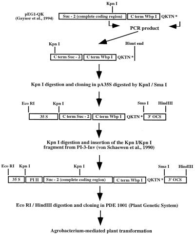
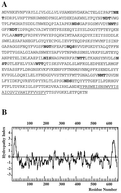
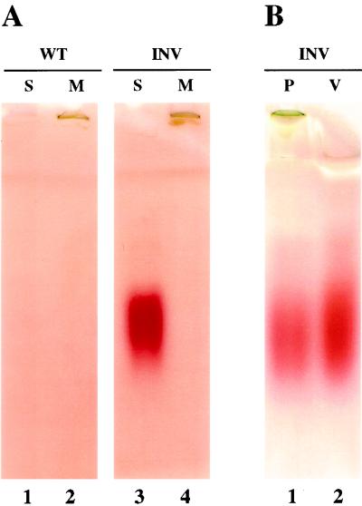
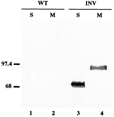
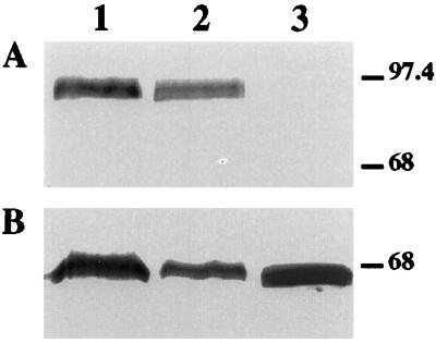
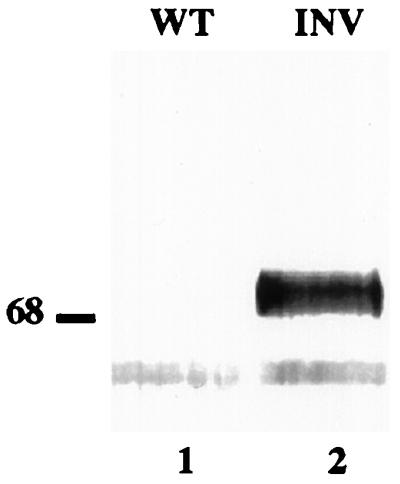
To further understand how membrane proteins are sorted in the secretory system, we devised a strategy that involves the expression of a membrane-anchored yeast invertase in transgenic plants. The construct consisted of a signal peptide followed by the coding region of yeast invertase and the transmembrane domain and cytoplasmic tail of calnexin. The substitution of a lysine near the C terminus of calnexin with a glutamic acid residue ensured progression through the secretory system rather than retention in or return to the endoplasmic reticulum. In the transformed plants, invertase activity and a 70-kD cross-reacting protein were found in the vacuoles. This yeast invertase had plant-specific complex glycans, indicating that transport to the vacuole was mediated by the Golgi apparatus. The microsomal fraction contained a membrane-anchored 90-kD cross-reacting polypeptide, but was devoid of invertase activity. Our results indicate that this membrane-anchored protein proceeds in the secretory system beyond the point where soluble proteins are sorted for secretion, and is detached from its membrane anchor either just before or just after delivery to the vacuole.


Figures

Figure 1
Figure 1
Summary of the construction of the plasmid used in this study.
Figure 2
Figure 2
Amino acid sequence and derived hydropathy plot of the membrane-anchored chimeric yeast invertase. A, Amino acid sequence of the fusion protein. The single-letter amino acid code is used. The signal peptide of the potato PR1 protein is represented in italics. The transmembrane domain of the yeast calnexin Wbp1 is underlined. Putative N-glycosylation sites are bolded. B, Hydropathy plot of the fusion protein. The plot was generated using a moving window of 11 residues (Kyte and Doolittle, 1982).
Figure 3
Figure 3
Detection of invertase activity in transgenic plants. A, Proteins from soluble (S, lanes 1 and 3) and microsomal (M, lanes 2 and 4) fractions of wild-type (WT) and transgenic (INV) leaves of transgenic plants were prepared as described in Methods and assayed for invertase activity after gel electrophoresis. Yeast invertase activity was detected only in the soluble fraction of transgenic plants. No invertase activity was detected in the soluble or microsomal fractions from wild-type plants. B, Detection of invertase activity in protoplasts (lane P) and vacuoles (lane V) of transgenic plants. Vacuoles were isolated from leaf protoplasts of transgenic plants and assayed for α-mannosidase activity. The invertase activity in the vacuole (lane 2) was compared with the invertase activity present in intact protoplasts (lane 1) after loading the same amount of α-mannosidase activity onto each lane.
Figure 4
Figure 4
Immunodetection of yeast invertase in protein extracts from transgenic plants. Proteins from soluble (S, lanes 1 and 3) and microsomal (M, lanes 2 and 4) fractions of wild-type (WT) and transgenic (INV) plants were fractionated by SDS-PAGE, electroblotted onto nitrocellulose membrane, and probed with a yeast invertase antiserum. Molecular standards are shown on the left (in kD).
Figure 5
Figure 5
Immunodetection of yeast invertase in microsomes of transgenic plants. A microsomal extract prepared from leaves of transgenic plants was subjected to five freeze-thaw cycles. After centrifugation at 100,000g for 1 h, proteins from the supernatant (soluble microsomal proteins, lane 3), the pellet (microsomal membrane proteins, lane 2) and the original total microsome fraction (lane 1) were separated by SDS-PAGE and blotted onto a nitrocellulose membrane. A, Immunodetection of yeast invertase. B, Immunodetection of BiP using a serum against tomato BiP. Molecular standards (in kD) are shown on the right.
Figure 6
Figure 6
Immunoblot analysis of proteins from the soluble fractions of wild-type and transgenic plants. Proteins from the soluble fractions of wild-type (WT, lane 1) and transgenic (INV, lane 2) plants were selectively immunoprecipitated using the yeast invertase antiserum, separated by SDS-PAGE, electroblotted onto nitrocellulose membrane, and probed with a plant complex glycan antiserum (Lauriere et al., 1989). Molecular standards are shown on the left (in kD).
Figure 7
Figure 7
Schematic diagram showing protein sorting in the secretory system. Membrane-anchored invertase reaches a compartment beyond the TGN, such as the PVC or the vacuole itself, before it is detached from the membrane. Whether invertase detachment from its membrane anchor occurs in the PVC or in the vacuole is not known. ○, Invertase.

References

    1. Ahmed SU, Bar-Peled M, Raikhel NV. Cloning and subcellular location of an Arabidopsis receptor-like protein that shares common features with protein-sorting receptors of eukaryotic cells. Plant Physiol. 1997;114:325–336. - PMC - PubMed
    1. Beevers L, Raikhel NV. Transport to the vacuole: receptors and trans elements. J Exp Bot. 1998;49:1271–1279.
    1. Boller T, Kende H. Hydrolytic enzymes in the central vacuole of plant cells. Plant Physiol. 1979;63:1123–1132. - PMC - PubMed
    1. Butcher H, Wagner GJ, Siegelman HW. Localization of acid hydrolases in protoplasts. Plant Physiol. 1977;59:1098–1103. - PMC - PubMed
    1. Chang A, Fink GR. Targeting of the yeast plasma membrane [H+]ATPase: a novel gene AST1 prevents mislocalization of mutant ATPase to the vacuole. J Cell Biol. 1995;128:39–49. - PMC - PubMed

Publication types

MeSH terms

LinkOut - more resources