. 1999 Aug;120(4):993-1004.
doi: 10.1104/pp.120.4.993.

Genetic and biochemical evidence for the involvement of alpha-1,4 glucanotransferases in amylopectin synthesis

Affiliations

Genetic and biochemical evidence for the involvement of alpha-1,4 glucanotransferases in amylopectin synthesis

C Colleoni et al. Plant Physiol. 1999 Aug.

Abstract

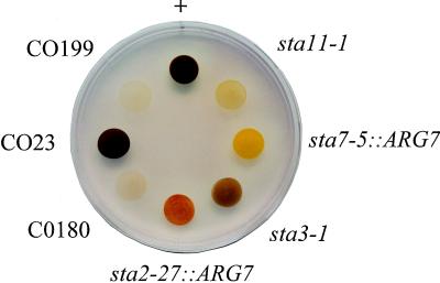
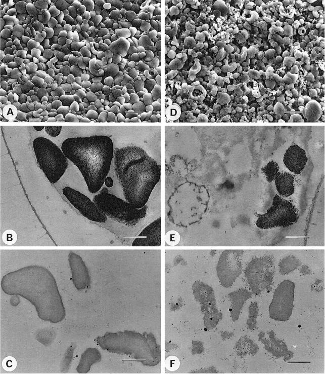
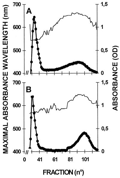
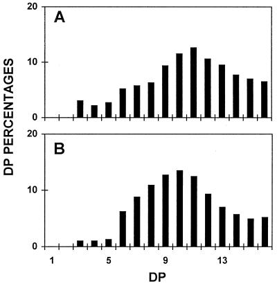
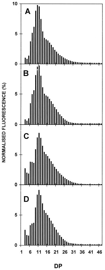
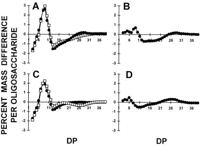
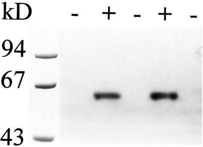
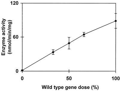
We describe a novel mutation in the Chlamydomonas reinhardtii STA11 gene, which results in significantly reduced granular starch deposition and major modifications in amylopectin structure and granule shape. This defect simultaneously leads to the accumulation of linear malto-oligosaccharides. The sta11-1 mutation causes the absence of an alpha-1,4 glucanotransferase known as disproportionating enzyme (D-enzyme). D-enzyme activity was found to be correlated with the amount of wild-type allele doses in gene dosage experiments. All other enzymes involved in starch biosynthesis, including ADP-glucose pyrophosphorylase, debranching enzymes, soluble and granule-bound starch synthases, branching enzymes, phosphorylases, alpha-glucosidases (maltases), and amylases, were unaffected by the mutation. These data indicate that the D-enzyme is required for normal starch granule biogenesis in the monocellular alga C. reinhardtii.


Figures

Figure 1
Figure 1
Wild-type and mutant iodine-staining phenotype. Iodine stain of cell patches incubated for 7 d on solid nitrogen-deprived medium. Genotypes with respect to starch are indicated for our reference strains. sta2–27::ARG7, sta3–1, and sta7–5::ARG7 correspond to highly specific defects respectively in GBSS, SS, and debranching enzyme (Delrue et al., 1992; Fontaine et al., 1993, Mouille et al., 1996). +, Wild type. The original mutant strain JV45J (sta11-1) and two recombinants, CO199 and CO180, display the typical yellow stain of low-starch mutants, while the wild-type recombinant CO23 shows the typical dark-blue color of the wild-type reference strain.
Figure 2
Figure 2
Growth curves of wild-type and mutant sta11-1 strains. Unsynchronized precultures of three mutant and three wild-type progeny from a cross involving the mutant JV45J and the wild-type strain 37 were grown in TAP medium (Harris, 1989b) to late log phase at 2 × 10⁶ cells mL−1. The cultures were inoculated at 5 × 10⁴ cells mL−1 and subjected to a 12-h d (80 μE m−2 s−1)/12-h night cycle of culture at 20°C with vigorous shaking. A, TAP medium (with acetate). Average mutant cell counts (▪) and wild-type (♦) cell counts are displayed as log of cell number versus time. B, TP medium (without acetate). Average mutant cell counts (▪) and wild-type (♦) cell counts are displayed as log of cell number versus time. The cultures are severely CO2 limited under these conditions and therefore grew at a very slow but comparable rate.
Figure 3
Figure 3
Relative frequency distributions of oligosaccharides. Undebranched malto-oligosaccharides accumulated by the sta11-1 reference strain JV45J were separated according to length after APTS fluorescence labeling and separation on a DNA sequencer. Percentages of chains ranging between a DP of 1 to 16 (chains containing 1–16 Glc residues) are scaled on the y axis.
Figure 4
Figure 4
1H-NMR analysis of malto-oligosaccharides. Part of the 1H-NMR spectra of amylopectin in dimethyl-sulfoxyde-d6/2H2O (80:20) at 80°C is displayed. The chemical shifts for the α- and β-anomers of the reducing end are at 5.1 and 4.5 ppm, respectively. If present, α-1,6-linkage anomeric protons should be found at 4.85 ppm. The integration area of the α- and β-anomers reducing end signals divided by that of the α-1,4-linkage anomeric proton at 5.2 ppm yields the average DP of the sample. NMR conditions were similar to those described in Delrue et al. (1992). A, Malto-oligosaccharides purified from strain JV45J. B, Maltotriose reference.
Figure 5
Figure 5
SEM and TEM of starches from wild-type and mutant sta11-1 strains. Electron micrographs of purified starches and in cells from nitrogen-starved wild-type (137C) and mutant sta11-1 (JV45J) C. reinhardtii strains. A to C, Wild-type strain 137C; D to F, mutant sta11-1 (JV45J) strain. A and D, SEM of purified starches (bar = 2 μm); B and E, TEM of starch-containing cells after PATAg staining (bar = 0.5 μm); C and F, TEM of purified starches after PATAg staining (bar = 0.5 μm).
Figure 6
Figure 6
Separation of amylopectin and amylose by CL2B-Sepharose chromatography. The optical density (•) was measured for each 2-mL fraction at λmax (unbroken thin line). All samples were loaded on the same column setup described by Delrue et al. (1992). The wild-type haploid 137C strain starch extracted from nitrogen-starved cultures (storage starch) (A) displays both amylopectin and low-Mr amylose. B, Starch from the mutant strain JV45J carrying the sta11-1 mutation. Starch was also extracted under nitrogen starvation (storage starch). Quantification of amylose and amylopectin ratios was obtained by pooling amylopectin and amylose fractions separately and measuring the amount of Glc through the standard amyloglucosidase assay.
Figure 7
Figure 7
Chain-length distributions of wild-type and mutant amylopectin. Isoamylase-debranched chains were separated according to length after APTS fluorescence labeling and separation on a DNA sequencer. Percentages of chains ranging between DP 1 to 16 (chains containing 1–16 Glc residues) are scaled on the y axis. A, Debranched chains of gel permeation chromatography-purified amylopectin from the mutant JV45J. B, Debranched chains from gel permeation chromatography-purified C. reinhardtii reference amylopectin (extracted from the amylose-free BAFR1 strain).
Figure 8
Figure 8
Chain-length distributions of amylopectin from wild-type and mutant strains. Distribution of chain lengths of wild-type and mutant amylopectin after isoamylase-mediated debranching were confirmed by capillary electrophoresis of APTS-labeled glucans following a procedure previously described (O'Shea et al., 1998). The relative amount of chains corresponding to each DP is strictly equivalent to the normalized fluorescence percentage. A and B correspond to wild-type strains 137C (A) and CO65 (B), while C and D correspond to sta11-1-carrying strains JV45J (C) and CO29 (D).
Figure 9
Figure 9
Comparison of the normalized masses of APTS-labeled oligosaccharides from isoamylase debranched amylopectin. The percentage difference of the total mass present in each individual oligosaccharide has been obtained by subtracting the chain-length distribution from debranched amylopectins. A, Subtractive analysis from the wild-type reference strain 137C minus that of the mutant sta11-1 JV45J (□) and from the wild-type strain 137C minus that of the mutant sta11-1 CO29 (•). B, Subtractive analysis from the wild-type reference strain 137C minus that of the wild-type strain CO65 (•). C, Subtractive analysis from the wild-type reference strain CO65 minus that of the mutant sta11-1 JV45J (□) and from the wild-type strain CO65 minus that of the mutant sta11-1 CO29 (•). D, Subtractive analysis from the mutant reference strain JV45J minus that of the mutant CO29 (•).
Figure 10
Figure 10
The enzymatic defect of sta11-1 mutant strains. Denatured crude extracts (100 μg of protein) from two wild-type (+) and three sta11-1 (−) were loaded on denaturing polyacrylamide gels. The proteins were renatured after electrophoresis and incubated overnight. Glc production was revealed after overnight incubation of the gel with 3 mg mL−1 maltotriose. The 62-kD blue-staining band can be easily distinguished in the wild-type cells. This band contained the D-enzyme activity.
Figure 11
Figure 11
Gene dosages expressed from 0% (homozygous mutant) to 100% (homozygous wild-type); 50% corresponds to the heterozygous diploids, while 33% and 66% correspond to a sta11-1/sta11-1/+ and a sta11-1/+/+ triploid respectively. Haploid, diploid, or triploid homozygous combinations gave identical results when the activity was expressed per milligram of protein. Means (♦) and sds of measures from three different diploid or triploid constructs were calculated for each gene dose. Total phosphoglucomutase-specific activities were monitored as internal controls and proved similar in all constructs (2.5 ± 0.4 nmol Glc-6-P formed from Glc-1-P min−1 mg−1 protein).

References

    1. Ball S. Regulation of starch biosynthesis. In: Rochaix J-D, Goldschmidt-Clermont M, Merchant S, Govindjee, editors. The Molecular Biology of Chloroplasts and Mitochondria in Chlamydomonas. Advances in Photosynthesis, Vol 7. Dordrecht, The Netherlands: Kluwer Academic Publishers; 1998. pp. 549–567.
    1. Ball S, Dirick L, Decq A, Martiat JC, Matagne RF. Plant Sci. 1990;66:1–9.
    1. Ball S, Guan H-P, James M, Myers A, Keeling P, Mouille G, Buléon A, Colonna P, Preiss J. From glycogen to amylopectin: a model explaining the biogenesis of the plant starch granule. Cell. 1996;86:349–352. - PubMed
    1. Ball S, Marianne T, Dirick L, Fresnoy M, Delrue B, Decq A. A Chlamydomonas reinhardtii low-starch mutant is defective for 3-phosphoglycerate activation and orthophosphate inhibition of ADP-glucose pyrophosphorylase. Planta. 1991;185:17–26. - PubMed
    1. Boos W, Shuman H. Maltose/maltodextrin system of Escherichia coli: transport, metabolism and regulation. Microbiol Mol Biol Rev. 1998;62:204–229. - PMC - PubMed

LinkOut - more resources