Human T lymphocyte priming in vitro by haptenated autologous dendritic cells
- PMID: 10444249
- PMCID: PMC1905350
- DOI: 10.1046/j.1365-2249.1999.00958.x
Human T lymphocyte priming in vitro by haptenated autologous dendritic cells
Abstract
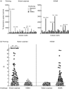
Dendritic cells (DC), generated from adherent peripheral blood mononuclear cells (PBMC) by culturing with granulocyte-macrophage colony-stimulating factor (GM-CSF) and IL-4, were used to study in vitro sensitization of naive, hapten-specific T cells and to analyse cross-reactivities to related compounds. DC were hapten-derivatized with nickel sulphate (Ni) or 2-hydroxyethyl-methacrylate (HEMA), followed by tumour necrosis factor-alpha (TNF-alpha)-induced maturation, before autologous T cells and a cytokine cocktail of IL-1beta, IL-2 and IL-7 were added. After T cell priming for 7 days, wells were split and challenged for another 7 days with Ni or HEMA, and potentially cross-reactive haptens. Hapten-specificity of in vitro priming was demonstrated by proliferative responses to the haptens used for priming but not to the unrelated haptens. Highest priming efficiencies were obtained when both IL-4 and IL-12 were added to the cytokine supplement. Marked interferon-gamma (IFN-gamma) release (up to 4 ng/ml) was found when IL-12 was included in the cultures, whereas IL-5 release (up to 500 pg/ml) was observed after addition of IL-4 alone, or in combination with IL-12. Nickel-primed T cells showed frequent cross-reactivities with other metals closely positioned in the periodic table, i.e. palladium and copper, whereas HEMA-primed T cells showed distinct cross-reactivities with selected methacrylate congeners. Similar cross-reactivities are known to occur in allergic patients. Thus, in vitro T cell priming provides a promising tool for studying factors regulating cytokine synthesis, and cross-reactivity patterns of hapten-specific T cells.
Figures


Similar articles
-
Interferon-alpha disables dendritic cell precursors: dendritic cells derived from interferon-alpha-treated monocytes are defective in maturation and T-cell stimulation.Immunology. 2003 Sep;110(1):38-47. doi: 10.1046/j.1365-2567.2003.01702.x. Immunology. 2003. PMID: 12941139 Free PMC article.
-
In vitro human T cell sensitization to haptens by monocyte-derived dendritic cells.Toxicol In Vitro. 2000 Dec;14(6):517-22. doi: 10.1016/s0887-2333(00)00043-6. Toxicol In Vitro. 2000. PMID: 11033063
-
Cross-reactivity of human nickel-reactive T-lymphocyte clones with copper and palladium.J Invest Dermatol. 1995 Jul;105(1):92-5. doi: 10.1111/1523-1747.ep12313366. J Invest Dermatol. 1995. PMID: 7615984
-
Human T cell priming assay: depletion of peripheral blood lymphocytes in CD25(+) cells improves the in vitro detection of weak allergen-specific T cells.Exp Suppl. 2014;104:89-100. doi: 10.1007/978-3-0348-0726-5_7. Exp Suppl. 2014. PMID: 24214620 Review.
-
The molecular basis of metal recognition by T cells.J Invest Dermatol. 1994 Apr;102(4):398-401. doi: 10.1111/1523-1747.ep12372149. J Invest Dermatol. 1994. PMID: 8151118 Review.
Cited by
-
A blinded in vitro analysis of the intrinsic immunogenicity of hepatotoxic drugs: implications for preclinical risk assessment.Toxicol Sci. 2023 Dec 21;197(1):38-52. doi: 10.1093/toxsci/kfad101. Toxicol Sci. 2023. PMID: 37788119 Free PMC article.
-
The LLNA: A Brief Review of Recent Advances and Limitations.J Allergy (Cairo). 2011;2011:424203. doi: 10.1155/2011/424203. Epub 2011 Jun 16. J Allergy (Cairo). 2011. PMID: 21747867 Free PMC article.
-
Drug and Chemical Allergy: A Role for a Specific Naive T-Cell Repertoire?Front Immunol. 2021 Jun 29;12:653102. doi: 10.3389/fimmu.2021.653102. eCollection 2021. Front Immunol. 2021. PMID: 34267746 Free PMC article. Review.
-
Identification and Characterization of Circulating Naïve CD4+ and CD8+ T Cells Recognizing Nickel.Front Immunol. 2019 Jun 12;10:1331. doi: 10.3389/fimmu.2019.01331. eCollection 2019. Front Immunol. 2019. PMID: 31249573 Free PMC article.
-
T-cell recognition of chemicals, protein allergens and drugs: towards the development of in vitro assays.Cell Mol Life Sci. 2010 Dec;67(24):4171-84. doi: 10.1007/s00018-010-0495-3. Epub 2010 Aug 18. Cell Mol Life Sci. 2010. PMID: 20717835 Free PMC article. Review.
References
-
- Tamaki K, Fujiwara H, Katz SI. Hapten specific TNP-reactive cytotoxic effector cells using epidemrmal cells as targets. J Invest Dermatol. 1981;7:225–9. - PubMed
-
- Krasteva M, Péguet-Navarro J, Moulton C, Courtellemount P, Redziniak G, Schmitt D. In vitro primary sensitization of hapten-specific T cells by cultured human epidermal Langerhans cells —a screening predictive assay for contact sensitizers. Clin Exp Allergy. 1996;26:563–70. - PubMed
-
- Dai R, Streilein JW. Naive, hapten-specific human T lymphocytes are primed in vitro with derivatized blood mononuclear cells. J Invest Dermatol. 1998;110:29–33. - PubMed
Publication types
MeSH terms
Substances
LinkOut - more resources
Full Text Sources

