. 1999 Aug 1;13(15):1960-9.
doi: 10.1101/gad.13.15.1960.

A cell-counting factor regulating structure size in Dictyostelium

Affiliations

A cell-counting factor regulating structure size in Dictyostelium

D A Brock et al. Genes Dev. .

Abstract

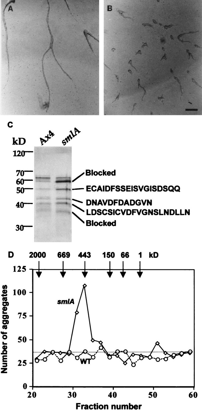
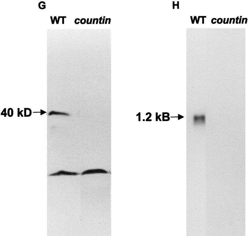
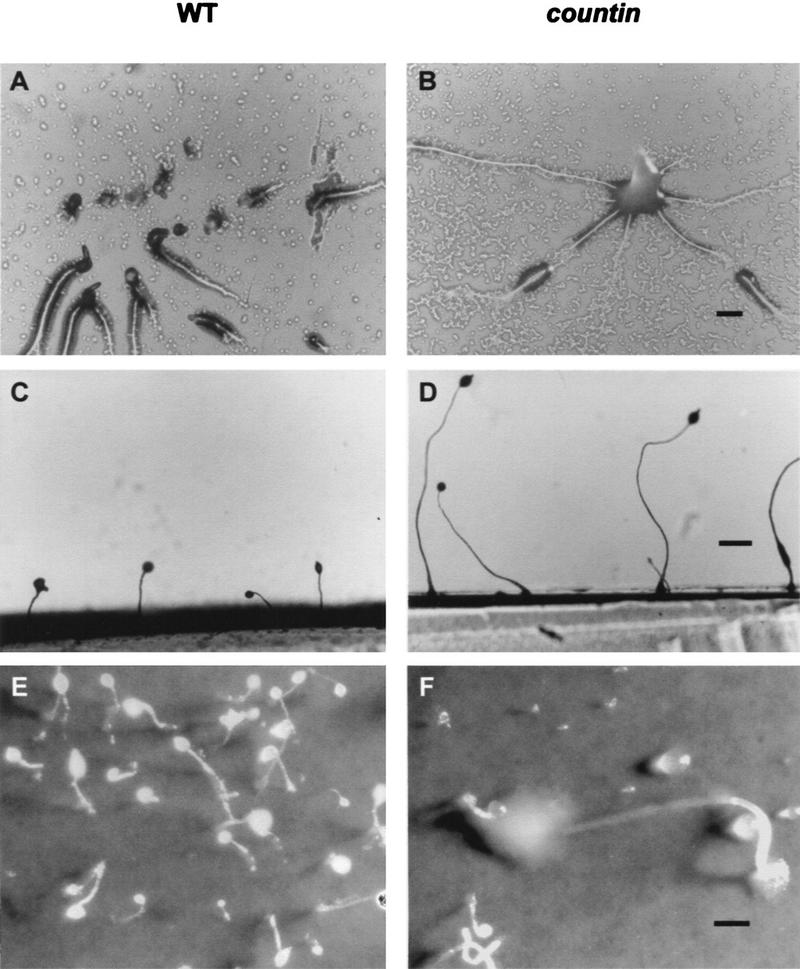
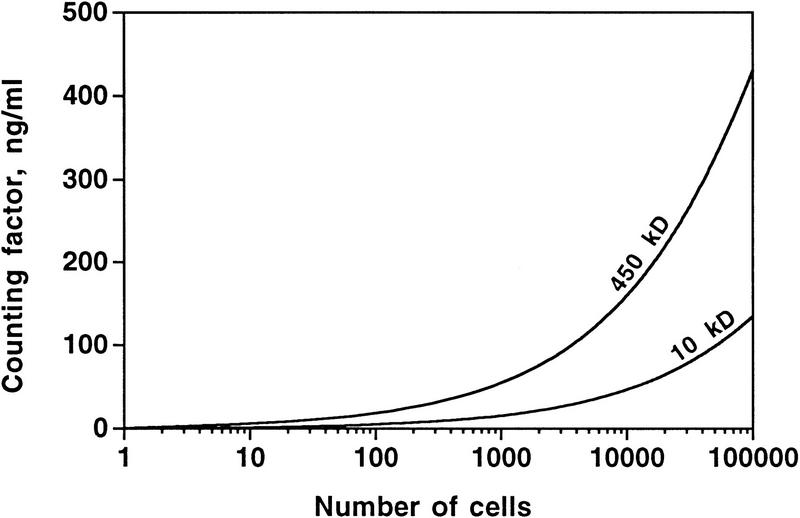
Developing Dictyostelium cells form large aggregation streams that break up into groups of 0.2 x 10(5) to 1 x 10(5) cells. Each group then becomes a fruiting body. smlA cells oversecrete an unknown factor that causes aggregation streams to break up into groups of approximately 5 x 10(3) cells and thus form very small fruiting bodies. We have purified the counting factor and find that it behaves as a complex of polypeptides with an effective molecular mass of 450 kD. One of the polypeptides is a 40-kD hydrophilic protein we have named counting. In transformants with a disrupted counting gene, there is no detectable secretion of counting factor, and the aggregation streams do not break up, resulting in huge (up to 2 x 10(5) cell) fruiting bodies.


Figures

Figure 1
Figure 1
Purification of the counting factor. (A,B) Bioassay of the purified counting factor activity. Fractions 18–23 from the hydroxyapatite column were pooled, concentrated, desalted, and run on a 10% nondenaturing polyacrylamide gel. Then, 0.5-cm slices were excised, electroeluted, and concentrated. Ax4 cells were starved in submerged culture for 15 hr in the presence of (A) the material eluted from slice 3 of the nondenaturing gel of Ax4 CM, (B) material eluted from the same slice from smlA CM. Slice 2 from the smlA preparation had a similar number of small aggregates as seen for slice 3 from smlA; all other slices from the smlA prep and all the slices from the Ax4 prep had normal aggregation streams as shown for the Ax4 slice 2. Bar in B, 200 μm. (C) Silver-stained SDS–polyacrylamide gel of the combined material eluted from gel slices 2 and 3. In other experiments, the polypeptide composition of the material from the two slices was seen to be identical. The amino-terminal amino acid sequences of the proteins are shown at right. The sequence we obtained for the 43-kD band was a mixture of two sequences; the sequence of the more abundant polypeptide is shown. (D) Gel-filtration chromatography of the active fractions from the hydroxylapatite column. (Solid line) Number of aggregates formed in the presence of fractionated smlA; (broken line) wild-type (WT) control; (dotted line) number of aggregates formed in PBM.
Figure 2
Figure 2
Sequence of the countin gene. The sequence starts with genomic DNA; the 5′ end of the cDNA is indicated by an arrow. The first AUG is at nucleotide 457. A broken underline indicates a potential signal sequence. The amino-terminal amino acid sequence obtained from the purified 40-kD protein is marked with a heavy underline. Two potential amino-linked glycosylation sites (Alexander 1997) are marked with boxes around the amino acid sequence. There is an intron between nucleotides 552 and 681. A light underline marks the sequence of the synthetic peptide used to immunize rabbits for antibody production. The KpnI sites used for the insertion of a blasticidin resistance cassette to make a gene disruption transformant are indicated by lines over the DNA sequence. (*) Start of an AAUAAA poly(A) addition signal. An open arrow marks where the cDNA sequence had 21 A’s and then terminated. The remainder of the sequence is genomic DNA. The sequence is available as AF140780 in Genbank.
Figure 3
Figure 3
Countin is present in all cells and is absent from countin transformants. (AF) Cells stained with anticountin antibody. (A,B,C) Immunofluorescence staining of cells; (D,E,F) corresponding phase images. A and D are Ax4 cells, B and E are countin knockout cells, and C and F are smlA cells. Bar in F, 10 μm. (G) Western blot of protein from wild-type (WT) and countin CM stained with anti-countin antibodies. (H) Northern blot of total RNA from vegetative cells probed with the countin cDNA.
Figure 3
Figure 3
Countin is present in all cells and is absent from countin transformants. (AF) Cells stained with anticountin antibody. (A,B,C) Immunofluorescence staining of cells; (D,E,F) corresponding phase images. A and D are Ax4 cells, B and E are countin knockout cells, and C and F are smlA cells. Bar in F, 10 μm. (G) Western blot of protein from wild-type (WT) and countin CM stained with anti-countin antibodies. (H) Northern blot of total RNA from vegetative cells probed with the countin cDNA.
Figure 4
Figure 4
The development of countin cells. (A,B) Aggregation of wild-type (WT) and countin cells. Cells were starved at 107 cells/cm2 on PBM agar. (A) Wild-type cells showing aggregation streams breaking up. (B) countin cells with unbroken streams forming a large aggregate. Bar, 200 μm. (C,D) Side views of wild-type and countin fruiting bodies. (C) Wild-type fruiting bodies are normal size. (D) Fruiting bodies formed by starved countin cells are much taller than wild-type. Bar, 500 μm. (E,F) The effect of forming large fruiting bodies. Cells were starved at 107 cells/cm2 on filter pads moistened with PBM. (E) A field of wild-type fruiting bodies depicting normal size. (F) A large, toppled countin fruiting body. Bar, 500 μm.
Figure 5
Figure 5
Assay of conditioned medium for counting factor activity. countin cells were starved in the presence of either PBM alone (Buffer), or buffer conditioned by a high density of starved countin, Ax4 (WT), or smlA cells. The number of aggregates was counted after 20 hr. Bars, s.e.m.
Figure 6
Figure 6
The theoretical concentration of counting factor at the center of a group of cells as a function of the number of cells in the group. The calculations were done for close-packed disks of cells on an agar or moist dirt surface, starting with one cell, then one cell with a ring of 6 cells around it (a total of 7 cells), up to a total of 200 concentric rings. The cells in the calculation were secreting counting factor continuously for 5 hr. The graph shows the concentrations for a 450-kD factor as well as a 10-kD factor, both being secreted from cells at 3 × 10−10 ng/sec.

References

    1. Alexander S. Developmental regulation and function of glycoproteins in Dictyostelium discoideum. In: Maeda Y, Inouye K, Takeuchi I, editors. Dictyostelium - A model system for cell and developmental biology. Tokyo, Japan: Universal Academy Press; 1997. pp. 349–362.
    1. Bonner JT, Hoffman ME. Evidence for a substance responsible for spacing pattern of aggregation and fruiting bodies in the cellular slime mold. J Embryol Exp Morphol. 1963;11:571–589. - PubMed
    1. Brock DA, Buczynski F, Spann TP, Wood SA, Cardelli J, Gomer RH. A Dictyostelium mutant with defective aggregate size determination. Development. 1996;122:2569–2578. - PubMed
    1. Clarke M, Gomer RH. PSF and CMF, autocrine factors that regulate gene expression during growth and early development of Dictyostelium. Experientia. 1995;51:1124–1134. - PubMed
    1. Compton T. Degenerate primers for DNA amplification. In: Innis MA, Gelfand DH, Sninsky JJ, White TJ, editors. PCR protocols. A guide to methods and applications. San Diego, CA: Academic Press; 1990. pp. 39–45.

Publication types

MeSH terms

Substances

Associated data