. 1999 Aug 1;13(15):1994-2004.
doi: 10.1101/gad.13.15.1994.

Uncoupling of the hnRNP Npl3p from mRNAs during the stress-induced block in mRNA export

Affiliations

Uncoupling of the hnRNP Npl3p from mRNAs during the stress-induced block in mRNA export

H Krebber et al. Genes Dev. .

Abstract

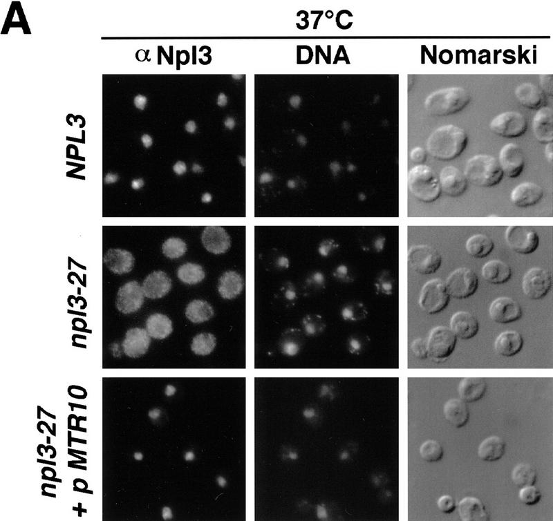
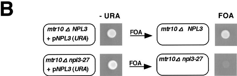
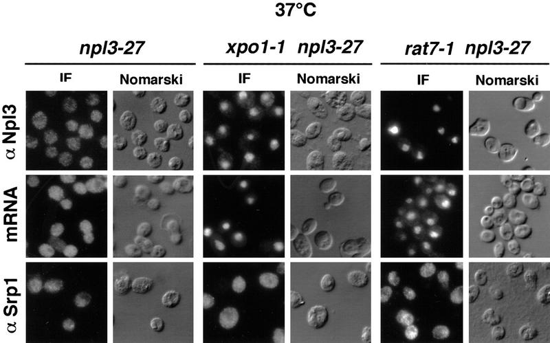
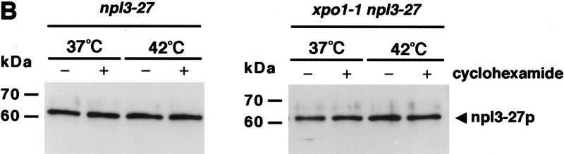
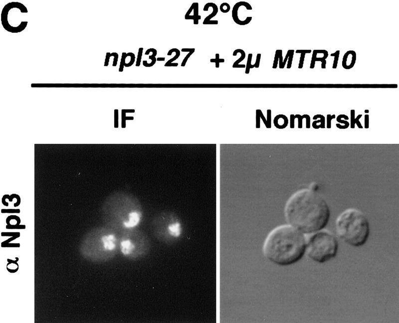
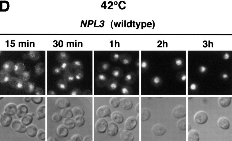
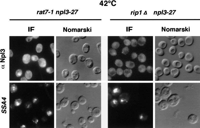
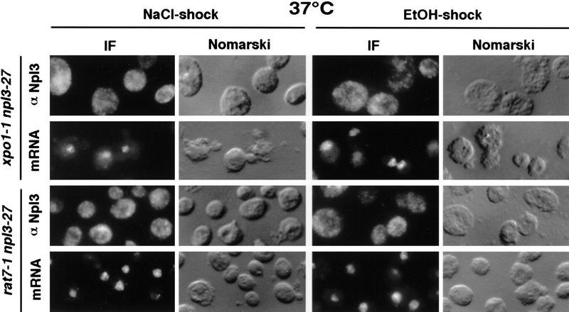
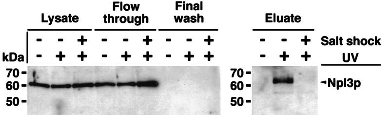
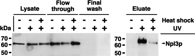
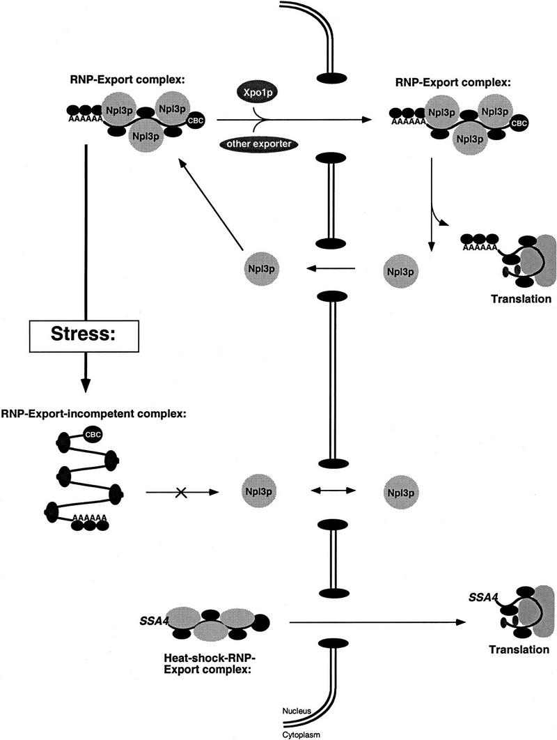
Npl3p, the major mRNA-binding protein of the yeast Saccharomyces cerevisiae shuttles between the nucleus and the cytoplasm. A single amino acid change in the carboxyl terminus of Npl3p (E409 --> K) renders the mutant protein largely cytoplasmic because of a delay in its import into the nucleus. This import defect can be reversed by increasing the intracellular concentration of Mtr10p, the nuclear import receptor for Npl3p. Conversely, using this mutant, we show that Npl3p and mRNA export out of the nucleus is significantly slowed in cells bearing mutations in XPO1/CRM1, which encodes the export receptor for NES-containing proteins and in RAT7, which encodes an essential nucleoporin. Interestingly, following induction of stress by heat shock, high salt, or ethanol, conditions under which most mRNA export is blocked, Npl3p is still exported from the nucleus. The stress-induced export of Npl3p is independent of both the activity of Xpo1p and the continued selective export of heat-shock mRNAs that occurs following stress. UV-cross-linking experiments show that Npl3p is bound to mRNA under normal conditions, but is no longer RNA associated in stressed cells. Taken together, we suggest that the uncoupling of Npl3p and possibly other mRNA-binding proteins from mRNAs in the nucleus provides a general switch that regulates mRNA export. By this model, under normal conditions Npl3p is a major component of an export-competent RNP complex. However, under conditions of stress, Npl3p no longer associates with the export complex, rendering it export incompetent and thus nuclear.


Figures

Figure 1
Figure 1
(A) Npl3-27p is slow in nuclear import. Wild-type (top), npl3-27 (middle), and npl3-27 cells overexpressing MTR10 on a 2μ plasmid (bottom) were grown to log phase and shifted to 37°C for 30 min. Npl3p localization was analyzed with an α-Npl3p antibody by immunofluorescence, the DNA was stained with DAPI, and the cells were photographed by Nomarski optics. (B) mtr10Δ and npl3-27 are synthetically lethal. The mtr10Δ npl3-27 double-mutant strain is not able to grow on a plate containing FOA, which allows only those cells to grow that are able to lose the URA3 plasmid (bottom, right). In the presence of wild-type NPL3 on a URA3 plasmid, cells are able to grow (bottom, left). Growth on FOA-containing medium is not impaired in the mutant mtr10Δ that contains genomic wild-type NPL3 (top).
Figure 1
Figure 1
(A) Npl3-27p is slow in nuclear import. Wild-type (top), npl3-27 (middle), and npl3-27 cells overexpressing MTR10 on a 2μ plasmid (bottom) were grown to log phase and shifted to 37°C for 30 min. Npl3p localization was analyzed with an α-Npl3p antibody by immunofluorescence, the DNA was stained with DAPI, and the cells were photographed by Nomarski optics. (B) mtr10Δ and npl3-27 are synthetically lethal. The mtr10Δ npl3-27 double-mutant strain is not able to grow on a plate containing FOA, which allows only those cells to grow that are able to lose the URA3 plasmid (bottom, right). In the presence of wild-type NPL3 on a URA3 plasmid, cells are able to grow (bottom, left). Growth on FOA-containing medium is not impaired in the mutant mtr10Δ that contains genomic wild-type NPL3 (top).
Figure 2
Figure 2
xpo1-1 and rat7-1 exhibit Npl3-27p localization and mRNA export defects. npl3-27 (left), xpo1-1 npl3-27 (middle), and rat7-1 npl3-27 (right) strains were grown to log phase then shifted to 37°C for 30 min before they were prepared for immunofluorescence (IF) with α-Npl3p (top), oligo[d(T50)] probe (middle), or α-Srp1p (bottom).
Figure 3
Figure 3
(A) At 42°C, Npl3-27p, but not mRNA exits the nucleus. Both strains, npl3-27 (left) and xpo1-1 npl3-27 (right), were grown to log phase, then shifted to 42°C for 15 min, before they were prepared for IF with α-Npl3p (top), oligo[d(T50)] probe (middle), and a SSA4 probe (bottom). (B) Yeast lysates of the same strains were prepared after they were grown to log phase and either incubated at 37°C for 3 hr (lanes 1,2) or to 42°C for 15 min (lanes 3,4) without [− (lanes 1,3)] or with cycloheximide addition [ + (lanes 2,4)]. The lysates were separated on a 12% SDS-polyacrylamide gel and analyzed on a Western blot with antibodies against Npl3p. (C) npl3-27 cells overexpressing MTR10 on a 2μ plasmid were grown to log phase and shifted to 42°C for 15 min. (D) Wild-type strains were grown to log phase before they were shifted to 42°C for different times. The localization of Npl3p was monitored by immunofluorescence.
Figure 3
Figure 3
(A) At 42°C, Npl3-27p, but not mRNA exits the nucleus. Both strains, npl3-27 (left) and xpo1-1 npl3-27 (right), were grown to log phase, then shifted to 42°C for 15 min, before they were prepared for IF with α-Npl3p (top), oligo[d(T50)] probe (middle), and a SSA4 probe (bottom). (B) Yeast lysates of the same strains were prepared after they were grown to log phase and either incubated at 37°C for 3 hr (lanes 1,2) or to 42°C for 15 min (lanes 3,4) without [− (lanes 1,3)] or with cycloheximide addition [ + (lanes 2,4)]. The lysates were separated on a 12% SDS-polyacrylamide gel and analyzed on a Western blot with antibodies against Npl3p. (C) npl3-27 cells overexpressing MTR10 on a 2μ plasmid were grown to log phase and shifted to 42°C for 15 min. (D) Wild-type strains were grown to log phase before they were shifted to 42°C for different times. The localization of Npl3p was monitored by immunofluorescence.
Figure 4
Figure 4
Npl3p does not exit the nucleus coupled to heat-shock mRNA. Both double mutants rat7-1 npl3-27 (left columns) and rip1Δ npl3-27 (right columns) were grown to log phase before they were shifted to 42°C for 15 min. Then they were analyzed by immunofluorescence with α-Npl3p (top) and by in situ hybridization with a SSA4 probe (bottom).
Figure 5
Figure 5
Salt- and ethanol shocks result in the export of Npl3-27p. Both double-mutant strains xpo1-1 npl3-27 (top two rows) and rat7-1 npl3-27 (bottom two rows) were grown to log phase and shifted to 37°C for 30 min before they were stressed with 0.4 m NaCl for 10 min (left) or with 10% ethanol for 30 min (right). Cells were then prepared for immunofluorescence (IF) with α-Npl3p (first and third row) and a dT probe (second and fourth row).
Figure 6
Figure 6
UV cross-linking of Npl3p to poly(A)+ RNA. (Top) Cells were either treated with or without high salt (+ or − Salt shock) and exposed to UV for cross-linking (+ or −UV-irradiation). The lysates were taken before application to the oligo(dT)–cellulose-column. (Flow through) The material that did not bind to the column. The final wash is derived from the last wash before elution (see Materials and Methods). The eluate contains the cross-linked poly(A)+ RNA-associated proteins. (Bottom) Cells were either shifted to 42°C for 15 min and exposed to UV (+heat shock +UV) or UV cross-linked without a heat shock (− heat shock +UV). The immunoblot was carried out with a polyclonal antibody against Npl3p.
Figure 6
Figure 6
UV cross-linking of Npl3p to poly(A)+ RNA. (Top) Cells were either treated with or without high salt (+ or − Salt shock) and exposed to UV for cross-linking (+ or −UV-irradiation). The lysates were taken before application to the oligo(dT)–cellulose-column. (Flow through) The material that did not bind to the column. The final wash is derived from the last wash before elution (see Materials and Methods). The eluate contains the cross-linked poly(A)+ RNA-associated proteins. (Bottom) Cells were either shifted to 42°C for 15 min and exposed to UV (+heat shock +UV) or UV cross-linked without a heat shock (− heat shock +UV). The immunoblot was carried out with a polyclonal antibody against Npl3p.
Figure 7
Figure 7
Model for RNP export under normal conditions and during stress. According to this model, mRNAs are packaged into export complexes by proteins such as Npl3p, which may interact with the as yet to be defined mRNA export machinery. Under conditions of stress, Npl3p dissociates from the RNP leaving it incompetent for export.

References

    1. Anderson JT, Wilson SM, Datar KV, Swanson MS. NAB2: A yeast nuclear polyadenylated RNA-binding protein essential for cell viability. Mol Cell Biol. 1993;13:2730–2741. - PMC - PubMed
    1. Amberg DC, Goldstein AL, Cole CN. Isolation and characterization of RAT1: An essential gene of Saccharomyces cerevisiae required for the efficient nucleocytoplasmic trafficking of mRNA. Genes & Dev. 1992;6:1173–1189. - PubMed
    1. Bogerd HP, Echarri A, Ross TM, Cullen BR. Inhibition of human immunodeficiency virus rev and human T-cell leukemia virus rex function, but not mason-pfizer monkey virus constitutive transport element activity, by a mutant human nucleoporin targeted to crm1. J Virol. 1998;72:8627–8635. - PMC - PubMed
    1. Bossie MA, DeHoratius C, Barcelo G, Silver P. A mutant nuclear protein with similarity to RNA binding proteins interferes with nuclear import in yeast. Mol Biol Cell. 1992;3:875–893. - PMC - PubMed
    1. Corbett AH, Silver PA. Nucleocytoplasmic transport of macromolecules. Microbiol Mol Biol Rev. 1997;61:193–211. - PMC - PubMed

Publication types

MeSH terms

Substances

LinkOut - more resources