. 1999 Jun-Jul;20(6):1015-24.

CNS involvement in neuro-Behçet syndrome: an MR study

Affiliations

CNS involvement in neuro-Behçet syndrome: an MR study

N Koçer et al. AJNR Am J Neuroradiol. 1999 Jun-Jul.

Abstract

Background and purpose: Behçet disease (BD) is a multisystem vasculitis of unknown origin in which neurologic involvement has been reported in the range of 5% to 10% in large series. Reports on clinical and radiologic aspects of neuro-Behçet syndrome (NBS) are in general limited in number. Our purpose was to determine the MR patterns in patients with NBS who had neural parenchymal involvement and to correlate our findings with possible vascular pathophysiology.

Methods: The MR images of 65 patients with NBS and neural parenchymal involvement were reviewed. In a subgroup of patients who had serial MR studies, we evaluated the anatomic-radiologic location and distribution of the lesions and whether they corresponded to any vascular territory, and studied their extension, enhancement patterns, and temporal course.

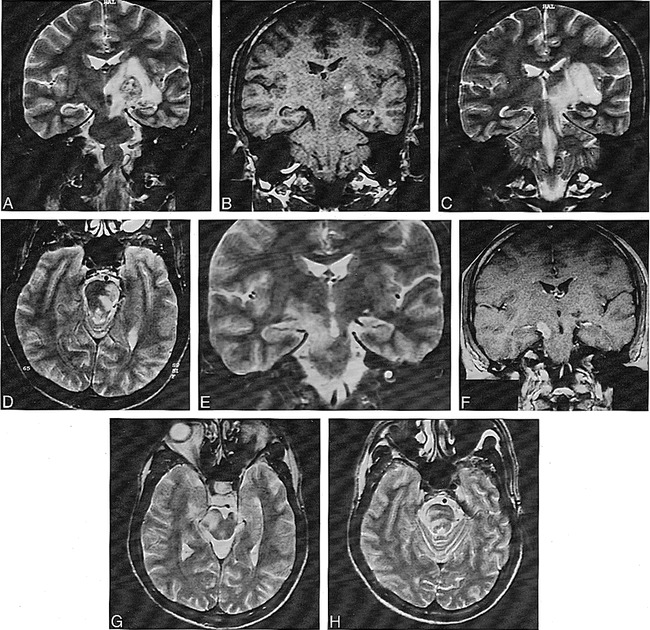
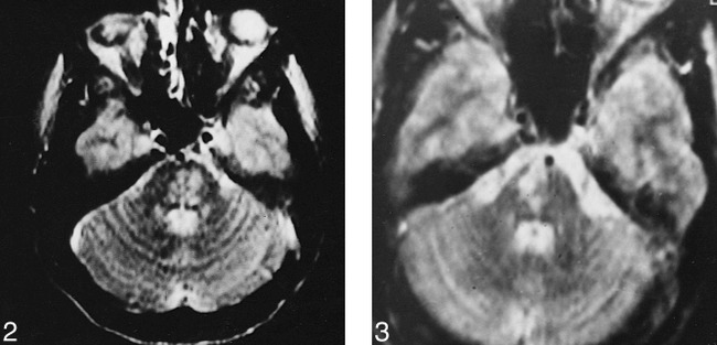
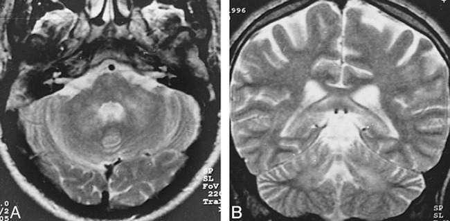
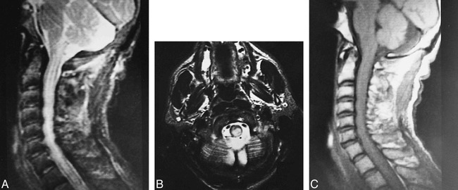
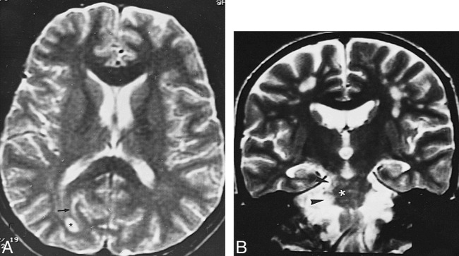
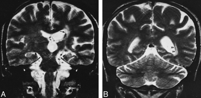
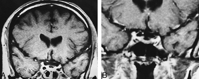
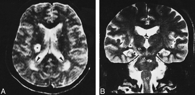
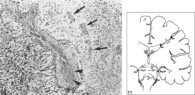
Results: The most common imaging finding in NBS patients who had neural parenchymal involvement was a mesodiencephalic junction lesion with edema extending along certain long tracts in the brain stem and diencephalon in 46% of the patients. The next most common location of involvement was the pontobulbar region, seen in 40% of the cases. Three primary cervical spinal cord lesions and one case of isolated optic nerve involvement were observed.

Conclusion: The parenchymal distribution of lesions in NBS appears to support the hypothesis of small-vessel vasculitis; mainly, venular involvement. The anatomic distribution of intraaxial veins of the CNS explains the predominant involvement of the brain stem structures observed in our patients. This pattern of lesion distribution might help to differentiate NBS from other vasculitides as well as from the inflammatory-demyelinating diseases of the CNS, such as multiple sclerosis.


Figures

<sc>fig</sc> 1.
fig 1.
A, Coronal T2-weighted image (4000/90/2 [TR/TE/excitations]) at the level of the crus cerebri nicely shows heterogeneous left MDJ lesion with extensive edema, sparing the red nucleus. B, Coronal noncontrast T1-weighted image (300/15/3) at the same level as A reveals a hemorrhagic focus in the lesion. C, Coronal T2-weighted image (4000/90/2) posterior to A shows extension of perilesional edema caudally to the superior cerebellar peduncle and pontine tegmentum, and upward to the white matter of the temporal lobe, external capsule, and thalamus. D, Caudal extension of the edema toward the pontine tegmentum is seen on axial T2-weighted image (4000/90/1). E, Two years later, after another relapse of the disease, coronal T2-weighted image (4000/90/1) reveals a contralateral MDJ lesion. The left-sided lesion now has shrunk to a small hypointense area. F, Contrast-enhanced T1-weighted image (660/17/1) shows enhancement of the new right MDJ lesion. G and H, Similar extension of edema as observed in C and D is seen in the right mesencephalopontine region of the brain stem on T2-weighted images (4000/90/1).
<sc>fig</sc> 2.
fig 2.
Axial T2-weighted image (2400/110/2) shows inhomogeneous hyperintense lesion located in the mid pons. fig 3. Axial T2-weighted image (2400/110/2) at the pontine level shows a right-sided pontine lesion that does not cross the midline.
<sc>fig</sc> 4.
fig 4.
A and B, Axial (A) and coronal (B) T2-weighted images (4000/90/2) reveal hyperintense lesions bilaterally in the middle cerebellar peduncles and deep cerebellar white matter
<sc>fig</sc> 5.
fig 5.
A–C, Midsagittal cervical lateral T2-weighted image (2200/80/1) (A) and axial T2-weighted image (4000/90/2) through the cervical medullary junction (B) show posteriorly located paracentral hyperintense lesion with mild cervicomedullary enlargement. The pattern of extension up to the inferior cerebellar peduncle suggests involvement of the dorsal columns. Midsagittal cervical lateral T1-weighted image (300/15/3) (C) shows slight enlargement of cervical cord
<sc>fig</sc> 6.
fig 6.
A, Axial T2-weighted image (3500/90/2) shows a well-defined deep right occipital white matter lesion (asterisk) and a subcortical linear hyperintensity (arrow). B, Coronal T2-weighted image (3500/90/1) in a different patient shows multiple subcortical white matter and right MDJ and pontine lesions (asterisk and arrowheads).
<sc>fig</sc> 7.
fig 7.
A, T2-weighted image (4000/90/2) shows chronic left MDJ lesion (double arrowhead) and ipsilateral lenticulostriatal lesions (arrow). B, Wallerian degeneration of the optic radiation is evident on paraatrial section of the same sequence (double arrowheads).
<sc>fig</sc> 8.
fig 8.
A, Contrast-enhanced coronal T1-weighted image (600/15/1) shows bilateral optic nerve atrophy and marked enhancement of the prechiasmatic segment of the right optic nerve (arrow). B, Contrast enhancement of the nerve disappeared after treatment (asterisk).
<sc>fig</sc> 9.
fig 9.
A, T2-weighted image (2400/110/2) shows round hyperintense lesion in the posterior and middle third of the right corona radiata (asterisk) associated with rather poorly defined periatrial T2 hyperintensities (arrowheads). B, Coronal T2-weighted image (4000/90/2) shows signal intensity changes along the right corticospinal tract, representative of secondary wallerian degeneration (double arrowheads), along with basal ganglia and pontine lesions (asterisks).
<sc>fig</sc> 10.
fig 10.
Histologic section of NBS shows a totally thrombosed medium-sized venous vessel. Focal fibrinoid necrosis (arrowheads) and moderate amount of lymphocyte and plasma cell infiltration is visible in the vessel wall. At the right side of the vessel, necrotic brain tissue with some newly thrombosed small vessel and mononuclear inflammatory infiltration (mainly lymphocytes and histiocytes) is seen (arrows). On the left, there is severe astrogliosis with some gemistocytic differentiation (H and E, original magnification ×100). fig 11. Schematic representation of the intraaxial venous system of normal parenchyma. Supratentorially, a medullary vein (1) permits bidirectional flow. In the brain stem, especially at the mesencephalic level, intraaxial veno-venous anastomosis (2) is sparse and venous flow is centrifugal, toward the pial veins (pv). ev indicates ependymal vein.

References

    1. Behçet H. Uber residivierende, aphtöse, durch ein virus verursachte Geschwüre am Mund, am Auge und an den Genitalien. Derm Woschenscr 1937;105:1152-1157
    1. Yazici H, Yurdakul S, Hamuryudan V. Behçet's syndrome. In: Klippel J, Dieppe P, eds. Rheumatology. London: Gower Medical; 1997;7.26:1–6
    1. O'Duffy JD. Vasculitis in Behçet's disease. Rheum Dis Clin North Am 1990;16:423-431 - PubMed
    1. Lakhanpal S, Tani K, Lie JT, Katoh K, Ishigatsuba Y, Ohokubo T. Pathological features of Behçet's syndrome: a review of Japanese autopsy registry data. Hum Pathol 1985;16:790-795 - PubMed
    1. Inaba G. Clinical features of neuro-Behçet syndrome. In: Lehner T, Barnes CG, eds. Recent Advances in Behçet's Disease. International Congress and Symposium Series Number 103. London: Royal Society of Medicine Services; 1986:235–246