

Adult-onset neurologic dysfunction associated with cortical malformations
- PMID: 10445440
- PMCID: PMC7056256
Adult-onset neurologic dysfunction associated with cortical malformations
Abstract
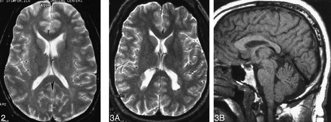
Background and purpose: Malformations of cerebral cortical development are common anomalies of the brain, typically causing developmental delay or seizures that are classically thought to begin in childhood. We present clinical and MR imaging data of 16 patients with cortical malformations in whom evidence of neurologic dysfunction was first noted in adulthood, and attempt to determine whether these malformations had any differentiating features from those presenting in childhood.
Methods: Imaging studies and clinical records of 16 patients with adult-onset neurologic dysfunction were reviewed retrospectively. The patients ranged in age from 17 to 64 years (mean age, 35 years) at the time of imaging. Imaging findings were correlated with seizure history.
Results: Fourteen patients had subependymal heterotopia (seven women, seven men), and two patients had closed-lip schizencephalies. Eleven patients had epilepsy, with age of onset ranging from 14 to 45 years (mean age, 22 years); four of them were successfully controlled by medication. The remaining five patients had no seizure disorder. All patients, except one, had normal intelligence. The bilaterality or multiplicity of location of heterotopias was not associated with the presence or absence of seizures, seizure frequency, or electroencephalographic results.
Conclusion: Subependymal heterotopia and small closed-lip schizencephaly may have minor clinical manifestations that are not evident until adulthood, or may, occasionally, never cause neurologic signs or symptoms whatsoever.
Figures


Similar articles
-
Subependymal heterotopia: a distinct neuronal migration disorder associated with epilepsy.J Neurol Neurosurg Psychiatry. 1994 Oct;57(10):1195-202. doi: 10.1136/jnnp.57.10.1195. J Neurol Neurosurg Psychiatry. 1994. PMID: 7931380 Free PMC article. Review.
-
Abnormalities of gyration, heterotopias, tuberous sclerosis, focal cortical dysplasia, microdysgenesis, dysembryoplastic neuroepithelial tumour and dysgenesis of the archicortex in epilepsy. Clinical, EEG and neuroimaging features in 100 adult patients.Brain. 1995 Jun;118 ( Pt 3):629-60. doi: 10.1093/brain/118.3.629. Brain. 1995. PMID: 7600083 Review.
-
Morphologic characteristics of subcortical heterotopia: MR imaging study.AJNR Am J Neuroradiol. 2000 Feb;21(2):290-5. AJNR Am J Neuroradiol. 2000. PMID: 10696010 Free PMC article.
-
Clinical features and long term outcome of epilepsy in periventricular nodular heterotopia. Simple compared with plus forms.J Neurol Neurosurg Psychiatry. 2004 Jun;75(6):873-8. doi: 10.1136/jnnp.2003.024315. J Neurol Neurosurg Psychiatry. 2004. PMID: 15146004 Free PMC article.
-
Subcortical heterotopia: a distinct clinicoradiologic entity.AJNR Am J Neuroradiol. 1996 Aug;17(7):1315-22. AJNR Am J Neuroradiol. 1996. PMID: 8871718 Free PMC article.
Cited by
-
Malformations of cortical development and epilepsy.Dialogues Clin Neurosci. 2008;10(1):47-62. doi: 10.31887/DCNS.2008.10.1/rjleventer. Dialogues Clin Neurosci. 2008. PMID: 18472484 Free PMC article. Review.
-
Magnetic resonance diffusion tensor imaging metrics in perilesional white matter among children with periventricular nodular gray matter heterotopia.Pediatr Radiol. 2013 Sep;43(9):1196-203. doi: 10.1007/s00247-013-2677-2. Epub 2013 Mar 26. Pediatr Radiol. 2013. PMID: 23529629
-
Malformations of cortical development: diagnostic accuracy of fetal MR imaging.Radiology. 2012 Jun;263(3):843-55. doi: 10.1148/radiol.12102492. Epub 2012 Apr 10. Radiology. 2012. PMID: 22495681 Free PMC article.
-
Adult-Onset Seizure Disorder Secondary to Schizencephaly.Asian J Neurosurg. 2020 Feb 25;15(1):159-161. doi: 10.4103/ajns.AJNS_293_19. eCollection 2020 Jan-Mar. Asian J Neurosurg. 2020. PMID: 32181192 Free PMC article.
-
Bipolar disorder with Melnick-Needles syndrome and periventricular nodular heterotopia: two case reports and a review of the literature.J Med Case Rep. 2021 Oct 11;15(1):495. doi: 10.1186/s13256-021-03064-1. J Med Case Rep. 2021. PMID: 34629090 Free PMC article. Review.
References
-
- Barkovich AJ, Chuang SH, Norman D. MR of neuronal migration anomalies. . AJNR Am J Neuroradiol 1987;8:1009-1017
-
- Barkovich AJ, Jackson DE, Boyer RS. Band heterotopias: a newly recognized neuronal migration anomaly. Radiology 1989;171:455-458 - PubMed
-
- Palmini A, Andermann F, Olivier A, et al. Neuronal migration disorders: a contribution of modern neuroimaging to the etiologic diagnosis of epilepsy. Can J Neurol Sci 1991;18:580-587 - PubMed
MeSH terms
LinkOut - more resources
Full Text Sources
Medical