

Case Reports
Partial midline fusion of the cerebellar hemispheres with vertical folia: a new cerebellar malformation?
Affiliations
- PMID: 10445461
- PMCID: PMC7056222
Item in Clipboard
Case Reports
Partial midline fusion of the cerebellar hemispheres with vertical folia: a new cerebellar malformation?
AJNR Am J Neuroradiol.
1999 Jun-Jul.
Abstract
MR imaging depicted vertically oriented folia instead of the normal horizontal folial pattern, hypoplastic cerebellar vermis, fusion of the inferior posterior cerebellum, and probable polymicrogyria in the superior cerebellar hemispheres in a child with hypotonia, nystagmus, ataxia, and psychomotor retardation. We propose that this newly discovered cerebellar malformation be added to the list of malformations associated with aplasia or hypoplasia of the cerebellar vermis, such as Dandy-Walker malformation, Joubert syndrome, tectocerebellar dysraphia, and rhombencephalosynapsis.
Figures
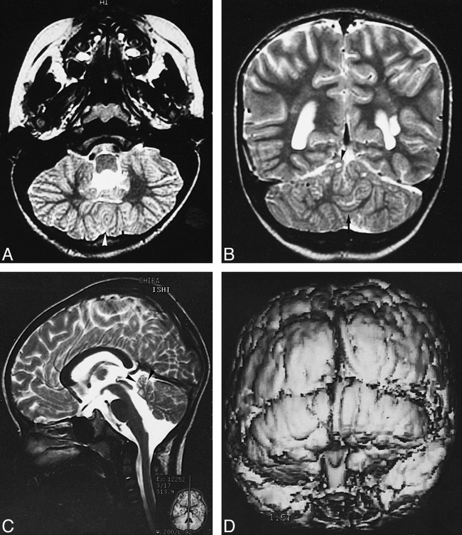
MR findings in a 4-year-old boy with hypotonia, nystagmus, ataxia, and psychomotor retardation. A, T2-weighted transverse image (3000/100/2 [TR/TE/excitations]) of the inferior cerebellum shows fusion of the cerebellar hemispheres (arrowhead) and dysplastic, vertically oriented, folial-like structures instead of the normally horizontal folia. B, Coronal T2-weighted image (3000/100/2) shows multifocal cystic lesions in the superior cerebellar hemispheres (arrowhead), a continuous thin layer of white matter (arrow), and a left periventricular leukomalacia. C, Sagittal T2-weighted image (3000/100/2) shows a small cerebellar vermis (arrowheads). D, Three-dimensional, fat-saturation, spoiled gradient-echo, surface shaded display image (50/6/1) shows vertically oriented folia.
References
-
- Barkovich AJ. Pediatric Neuroimaging.. 2nd ed. New York: Raven Press; 1995:246–257
-
- Ramaekers VT, Heimann G, Reul J, Thron A, Jaeken J. Genetic disorders and cerebellar structural abnormalities in childhood. Brain 1997;120:1739-1751 - PubMed
-
- Freide RL. Developmental Neuropathology.. 2nd ed. Berlin: Springer; 1989:361–371
-
- Maria BL, Hoang KB, Tusa RJ, et al. Joubert syndrome revisited: key ocular motor signs with magnetic resonance imaging correlation. J Child Neurol 1997;12:423-430 - PubMed
-
- Smith MT, Huntington HW. Inverse cerebellum and occipital encephalocele. Neurology 1977;27:246-251 - PubMed
Publication types
MeSH terms
LinkOut - more resources
Full Text Sources