

Review
doi: 10.1136/gut.45.3.317.
Hereditary pancreatitis: new insights into acute and chronic pancreatitis
Affiliations
- PMID: 10446089
- PMCID: PMC1727629
- DOI: 10.1136/gut.45.3.317
Item in Clipboard
Review
Hereditary pancreatitis: new insights into acute and chronic pancreatitis
Gut.
1999 Sep.
No abstract available
Figures
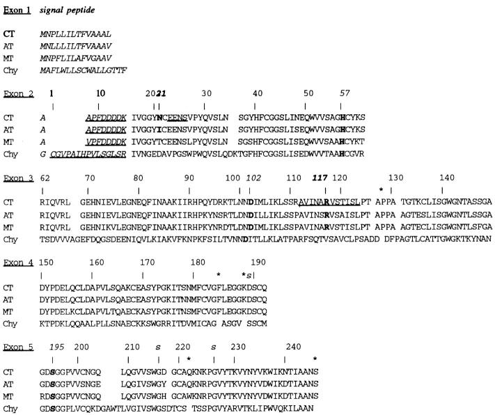
Cationic trypsinogen (CT) compared with anionic trypsinogen (AT), mesotrypsinogen (MT) and chymotrypsinogen (Chy). Cationic trypsinogen is divided according to the amino acids coded for in each of the five exons. The numbering system is based on alignment with chymotrypsinogen at serine 195.16 The sequences of cationic and anionic trypsinogen are from17; mesotrypsinogen from18 and19; and chymotrypsinogen from cDNA (introns unknown).20 Signal peptides are in italics, activation peptides are underlined italics , the catalytic triad (H57, D102 and S195), N21, and R117 are bold. The amino acids determining specificity of the enzymes (D189, G216 and G226) are marked with "s". Amino acids in trypsinogen that occur between the chymotrypsinogen numbered residues are marked with an asterisk.
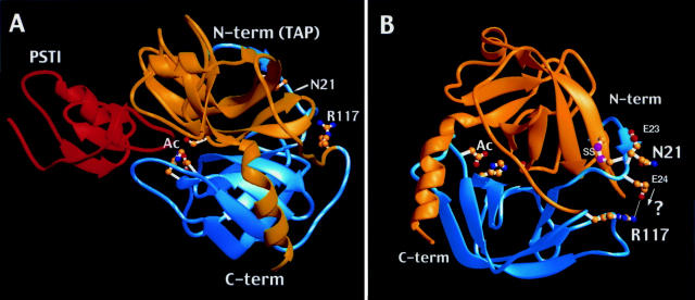
Crystallographic structure of trypsin. (A) The two domain structure of bovine trypsinogen-pancreatic secretory trypsin inhibitor (PSTI) complex.35 Note the transition from the blue N-terminal (N-term) domain to the yellow C-terminal (C-term) domain at R117 in the flexible chain segment connecting the domains. The trypsinogen activation peptide (TAP) portion of the molecule resides in the N-term region but is not visualised crystallographically. PSTI (red) is shown interacting with the active site (Ac) of trypsin. The location of N21 is also noted. (B) The two domain structure of human cationic trypsin.21 Structural location of N21, E24, and R117. One possible effect of the N21I substitution would be to bring E24 closer to R117, forming a salt bridge between the oppositely charged side chains (arrow and "?").
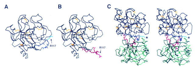
Limited proteolysis model of trypsin by trypsin. The backbone of the primary trypsinogen (black) is illustrated with the A112-L123 side chain shown in native conformation (A; blue) and the modelled conformation (B; red) that may be necessary for limited proteolysis of trypsin at R117 (arrow) by another trypsin molecule. (C) Stereo view of the trypsin-trypsin autolysis limited proteolysis model with a second trypsin (green) in the attack position. The two trypsin molecules are oriented with the active site facing upward. The A112-L123 side chain from A and B are both illustrated, with R117 in the native position marked with a asterisk. When the side chain is in the modelled (red) conformation the R117 of the primary (black) trypsin molecule easily fits within the active site of the second trypsin molecule. (To see the molecule in three dimensions, position the figure approximately 30 cm (12 inches) from the face, relaxing and crossing the eyes to allow the images to merge. One should see three images, with the one in the middle appearing in three dimensions.)
References
Publication types
MeSH terms
Substances
Grants and funding
LinkOut - more resources
Full Text Sources
Other Literature Sources
Medical